Rotator cuff calcific tendinopathy (RCCT) is a common condition caused by calcium deposits in the rotator cuff tendons or subacromial bursa. These calcific deposits are found in 3 to 20 percent of asymptomatic adults and in a substantially higher proportion of patients presenting with shoulder pain. The condition primarily affects adults aged 30 to 60 and is slightly more common in females. Calcific deposits can trigger inflammation or irritate the surrounding tissue and bursa, causing acute or chronic pain.
Initial treatment includes physical therapy, NSAIDs, activity modification, and often corticosteroid injections. If a patient’s pain persists, minimally invasive procedures such as ultrasound-guided needling and lavage (barbotage) can be effective. Some clinical trials have found greater deposit clearance and pain reduction with lavage compared with extracorporeal shockwave treatment (ESWT) or corticosteroid injection alone, though the quality of evidence has been mixed. Surgery is considered for patients with symptoms that remain disabling despite an adequate course of nonsurgical treatment.
Here we present a case of a healthy female with persistent left shoulder pain who was diagnosed with rotator cuff calcific tendinopathy and successfully treated with ultrasound-guided barbotage and subsequent aspiration.
Case Highlights:
- An active 43-year-old female presented with chronic left shoulder pain secondary to rotator cuff calcific tendinopathy.
- Two years of conservative treatment failed to provide lasting relief.
- The patient was treated with barbotage to resolve her persistent symptomatic calcific tendinopathy.
- To mitigate the subsequent risk of calcific debris migration into the bursa, ultrasound-guided aspiration was used to treat acute calcific bursitis.
- At a 2-year follow-up, the patient’s calcific deposits had completely resolved and she reported continued pain relief.
Case Presentation
A healthy 43-year-old woman presented to the outpatient sports medicine office in February 2020 with left shoulder pain that had persisted for 6 months. She did not have a specific injury or trauma but had been weightlifting more consistently. The patient reported feeling pain when putting on clothes and when sleeping on the affected shoulder.
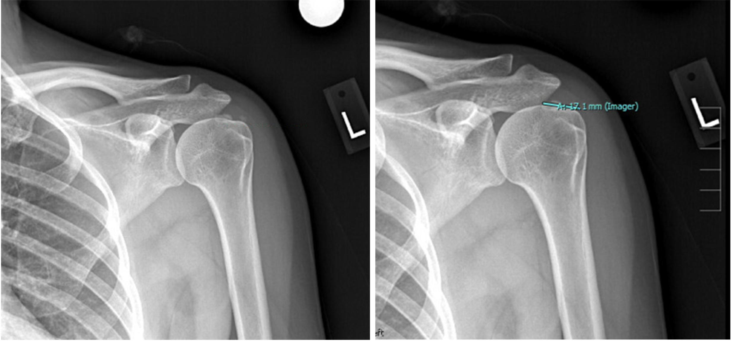
On examination, the patient’s pain was reproduced within 60 to 120 degrees of abduction and her left shoulder showed positive signs for impingement. An X-ray (Figure 1) was remarkable for a calcium deposit in the supraspinatus tendon and likely the infraspinatus tendon that measured 1.7 by 1.2 cm. The patient was referred to physical therapy for strengthening, which was limited due to shutdowns during the COVID-19 pandemic.
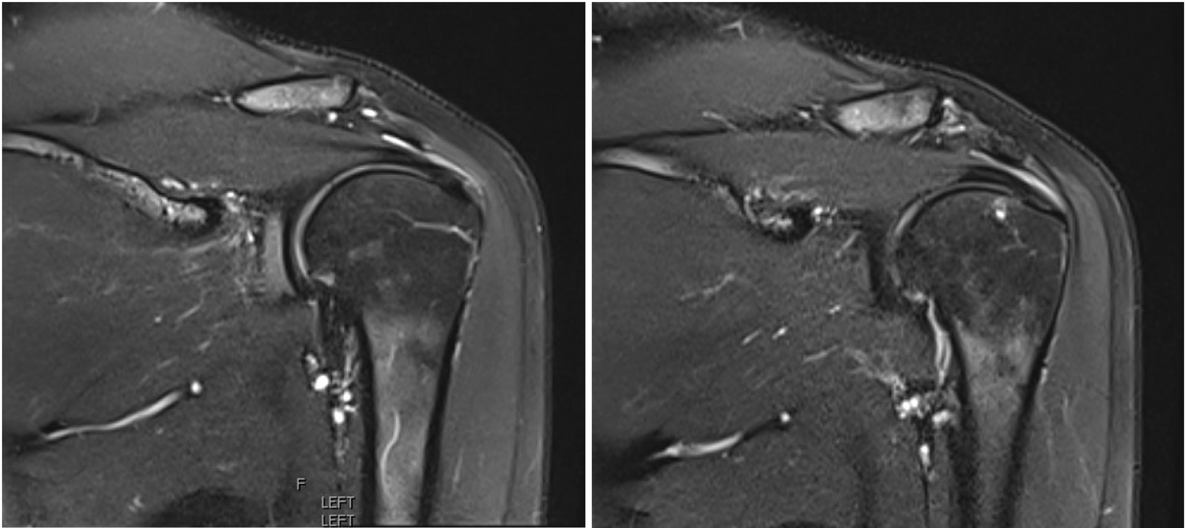
At a follow-up visit 4 months later in June 2020, the patient showed partial improvement, although she reported continued pain with her activities of daily living. Her exam was relatively unchanged and redemonstrated impingement and a 4/5 strength score with external rotation and forward elevation. An MRI of the left shoulder in July 2020 (Figure 2) showed a low-grade partial thickness intrasubstance tear of the posterior supraspinatous and anterior infraspinatous. In addition, it showed focal calcium hydroxyapatite deposition (measuring 1.1 by 0.6 cm) at the distal insertion of the posterior supraspinatous and anterior infraspinatous tendon as well as adjacent subacromial bursitis.
The woman was offered a subacromial corticosteroid injection (CSI) but declined and opted for additional physical therapy, which led to improvement but not a complete resolution of her symptoms.
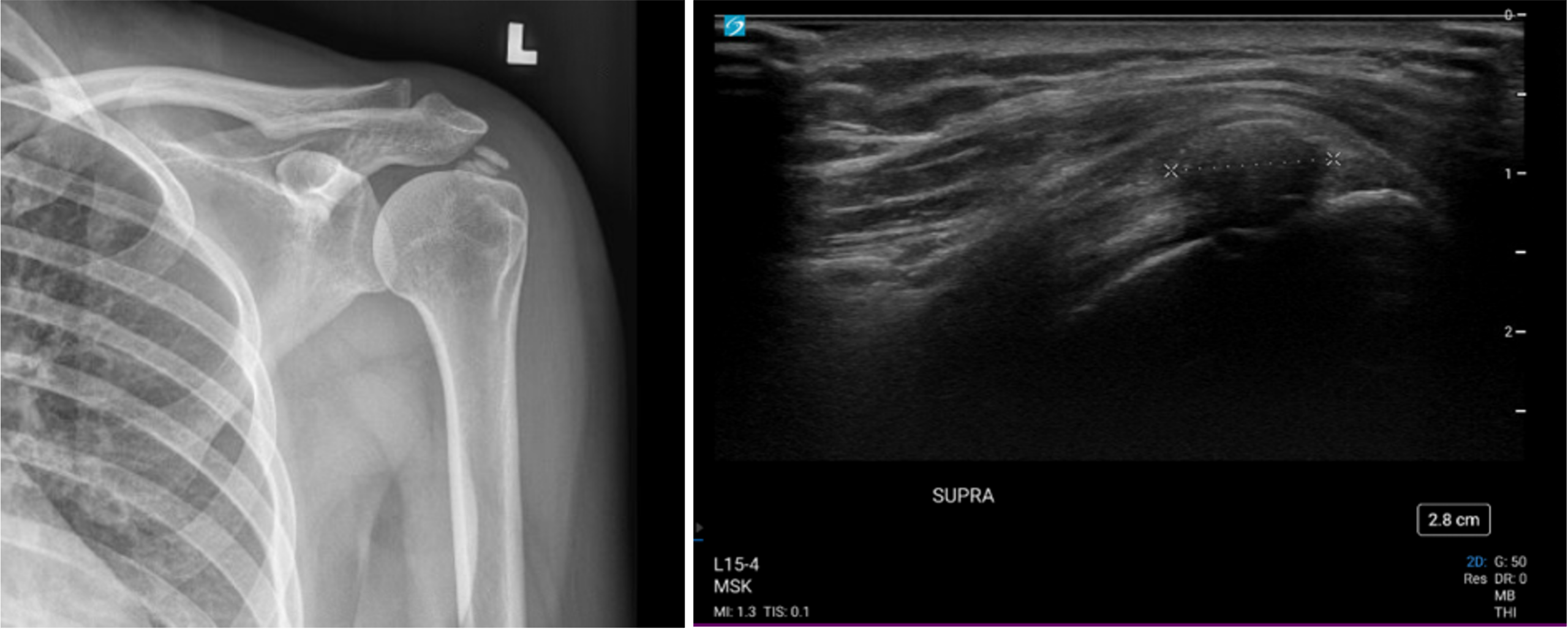
The patient subsequently presented with severe left shoulder pain in December 2023. She reported mild, intermittent pain since her last evaluation, with a recent exacerbation of that pain. On exam, her range of motion was limited in forward elevation compared to the contralateral side and the left shoulder impingement signs were again positive. Her strength score was 4+/5 with abduction and she reported significant pain with resisted abduction. The calcific deposits were larger than on the initial presentation and were redemonstrated within the supraspinatus on an X-ray of her left shoulder (Figure 3, left) and on bedside ultrasound (Figure 3, right).
The patient again opted for conservative treatment starting with physical therapy. She did not notice relief after 3 months of consistent treatment, however, and opted for barbotage without accompanying steroid treatment.
Nonsurgical Management
The patient was placed supine on the exam table with the head raised in a reclined position; her arm was internally rotated and slightly abducted. Ultrasound was used to locate the calcific deposits. The area was cleaned and prepped in standard fashion. The patient was given local anesthesia with 1 percent lidocaine to the skin and directly around the calcium deposits.
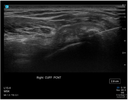
Under direct ultrasound guidance, an 18G needle was advanced to the largest deposit and multiple gentle needle fenestrations were made to assess the composition of the deposit, which was noted to be well-formed and bone-like. Using additional force, the needle was advanced into the core of the deposit and barbotage was performed, with repeated injection and aspiration of 0.5 to 1 mL of sterile saline (Figure 4, Video 1). The fenestration and barbotage were repeated with the goal of expanding each deposit and aspirating the calcific debris. Upon aspirating the debris, the syringe was exchanged for a new one. The patient tolerated the procedure well and her left arm was placed in a sling for 2 days post-procedure with daily mobilization exercises.
Source: NYU Langone Health
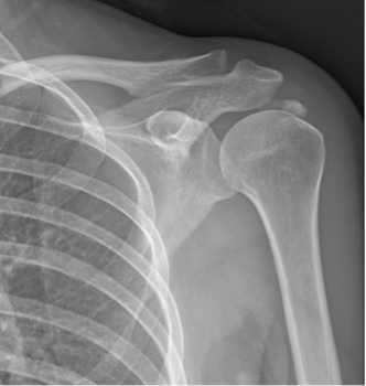
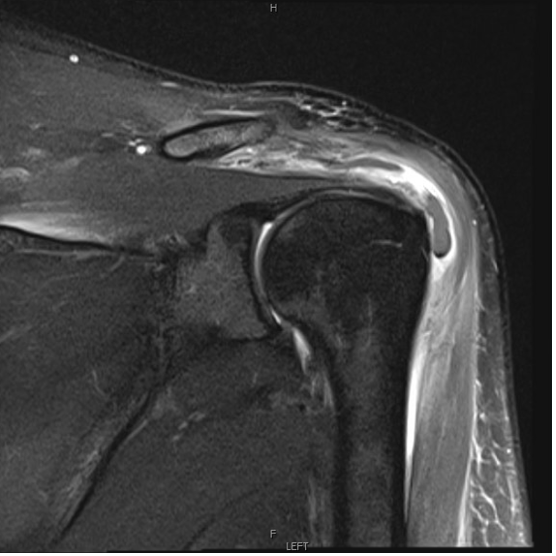
At a 2-week post-procedure follow-up visit, the patient reported improvement in her symptoms. Roughly 8 weeks after the procedure, however, she had a flare of acute severe pain and was evaluated at the Orthopedic Immediate Care Center, where an X-ray showed persistent albeit less homogenous calcific tendinopathy (Figure 5). She was referred for an MRI, which showed concern for acute subacromial/subdeltoid calcific bursitis and partial bursal side tearing of the anterior supraspinatus tendon (Figure 6).
Source: NYU Langone Health
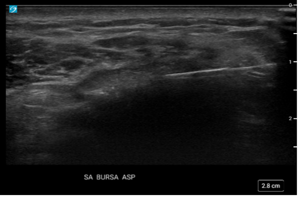
Following post-MRI treatment for the patient’s pain, an ultrasound-guided left subacromial subdeltoid bursa aspiration removed 2 mL of thick, white chalky fluid (Figure 7) while a repeat aspiration 3 days later removed an additional 2 mL of serosanguinous fluid. As a precaution, the fluid was sent for culture, and septic bursitis was ruled out as a cause of the patient’s acute pain. The aspiration provided her with some pain relief.
Source: NYU Langone Health
Post-Procedural Course
Given the patient’s persistent calcific tendinopathy on X-ray and the extent of her pain, she underwent a surgical consultation in May 2024, with a recommendation for surgical intervention with debridement (+/- cuff repair if needed). The patient deferred surgery due to travel plans, and continued NSAIDs for pain control and at-home physical therapy.
At a follow-up visit in August 2024, the patient reported that her pain had significantly improved. A follow-up X-ray showed resolution of the calcific tendinosis (Figure 8, left) and an MRI similarly showed no evidence of calcific deposits and improvement of the partial supraspinatus partial tearing, with a persistent low grade partial thickness tear of the infraspinatus (Figures 8, middle and right).
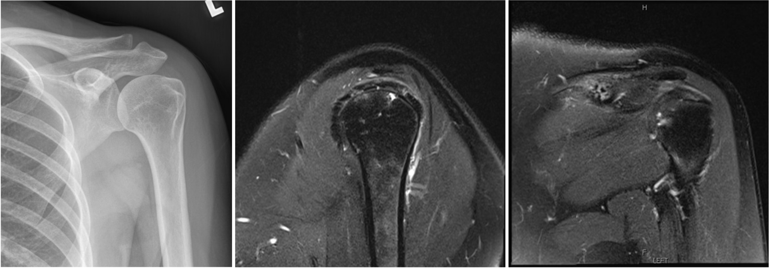
The patient continued her at-home physical therapy. At regular follow-up visits through early 2025 to evaluate her left shoulder, and at a visit in March 2026 for an unrelated complaint, she continued to have complete resolution of her shoulder pain.
Discussion
Calcific tendinopathy is a dynamic disease process rather than a static calcium deposit within the rotator cuff. The condition progresses through formative, resting, and resorptive phases, with spontaneous resorption occurring in some patients. However, symptomatic deposits may persist for years despite nonsurgical management, resulting in chronic pain and functional limitation. In such patients, treatment may be directed not only at symptom control but also at facilitating disruption and resorption of the calcific deposit.
Ultrasound-guided lavage, or barbotage, provides a minimally invasive method to directly target the calcific deposit. In some cases, mechanical disruption and debulking of the deposit may be sufficient to reduce the local mechanical and inflammatory effects of the calcium and lead to symptomatic improvement. However, barbotage may also stimulate the body’s natural resorptive process by fenestrating the solid deposit. This process may be particularly relevant in patients with longstanding calcium deposits that have failed to resolve spontaneously, as exemplified by our patient described here.
In the clinical course for our patient, she developed an acute episode of calcific subacromial bursitis approximately 2 months post-barbotage. Aspiration of the debris under ultrasound guidance led to gradual improvement in her symptoms over the subsequent 3 to 4 months, with eventual resolution of both her pain and the calcific tendinopathy. This case highlights the time it takes to achieve tissue healing and repair, which can help set expectations with patients. We were able to avoid a corticosteroid injection and surgical debridement in our patient, in accordance with her preference.
Extracorporeal shockwave therapy is another nonsurgical treatment option for chronic symptomatic calcific tendinopathy and is thought to promote fragmentation and resorption of calcific deposits. Clinical studies have demonstrated improvement in pain and function as well as changes in deposit size following shockwave therapy, although treatment protocols and outcomes vary. Percutaneous needle tenotomy, including ultrasonic techniques such as Tenex, provides another minimally invasive approach by mechanically disrupting and removing pathologic tendon tissue and may have a role in selected patients.
For patients with persistent symptomatic calcific tendinopathy, this case demonstrates the role of ultrasound-guided intervention in the office setting. In particular, barbotage represents a minimally invasive and effective treatment option that directly targets the underlying deposit while avoiding the need for surgical intervention.
References
- Kim MS, et al. Clin Shoulder Elb. 2020;23(4):210-216. DOI.
- Merolla G, et al. J Orthop Traumatol. 2016;17(1):7-14. DOI.
- Avendano JP, Pereira D. Orthopedics. 2023;46(6):e326-e332. DOI.
- de Witte PB, et al. Am J Sports Med. 2013;41(7):1665-1673. DOI.
- Sconza C, et al. Eur J Phys Rehabil Med. 2024;60(6):995-1008. DOI.
- van Noort DM, et al. Arthroscopy. 2026;42(11):2485-2493. DOI.