Case Highlights:
- Ultrasound and MRI imaging confirmed a recurrently draining and bleeding lymphovenous malformation in combination with a calvarial defect in a 5-week-old infant.
- Surgical resection of the involved skin and 90 percent of the cyst wall removed the mass and greatly reduced the risk of recurrence.
- The infant’s 1.2 cm calvarial defect was corrected, with an advancement flap procedure used to move unaffected scalp skin for incision closure.
A 5-week-old infant girl presented to Hassenfeld Children’s Hospital at NYU Langone Health in October 2025 with a 9 by 5.5 cm mass on her right posterior scalp that had been diagnosed via prenatal ultrasound. The mass had repeatedly leaked blood and fluid, especially after a frenulectomy performed shortly after her birth.
Although the baby had been feeding and gaining weight appropriately, her mother expressed concern over the “watch and wait” strategy adopted by another medical institution.
“This malformation was significant. I knew we needed to jump on it, that it wasn’t something to just watch.”
Francine Blei, MD
The family self-referred to pediatric hematologist-oncologist Francine Blei, MD, director of NYU Langone’s Hemangioma and Vascular Malformations Program. On examination, Dr. Blei noted that the infant’s compressible mass showed serosanguinous drainage transillumination (indicating a buildup of bloody proteinaceous fluid), multiple dermal channels, and peau d’orange changes.
Ultrasound suggested that the mass was due to a lymphatic malformation that was unlikely to resolve on its own, and Dr. Blei quickly alerted a team of pediatric specialists, including surgeon Sandra S. Tomita, MD, neurosurgeon David H. Harter, MD, and plastic surgeon Sheel Sharma, MD. “This malformation was significant,” Dr. Blei says. “I knew we needed to jump on it, that it wasn’t something to just watch.”
A skull irregularity suggested potential involvement with the brain, and further skin erosion raised the risk of a more substantial defect, additional bleeding, and an infection. “We all took that very seriously and recognized that we needed to fast-track this case,” she says.
An Unusual Congenital Combination
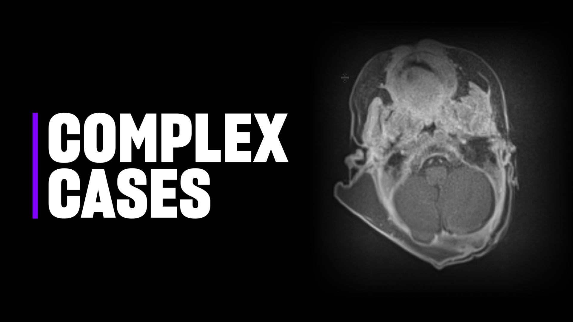
An MRI revealed a macrocystic right suboccipital lymphatic malformation with traversing veins in addition to a calvarial defect with associated encephalocele. “This was a very uncommon constellation of findings that I hadn’t seen before,” Dr. Harter says. “It required flexibility and good collaboration among our teams.”
Dr. Tomita led the effort to surgically resect the mass, with Dr. Sharma assisting. “The patient’s skin was also involved in the malformation, so we had to remove some of that skin,” Dr. Sharma says. Otherwise, the patient would have had a high risk of recurrence. The remaining lymphatic malformation involved deeper tissues; together, the surgeons excised roughly 90 percent of the cyst wall.
“This was a very uncommon constellation of findings that I hadn’t seen before. It required flexibility and good collaboration among our teams.”
David H. Harter, MD
Dr. Harter then corrected the infant’s 1.2 cm calvarial defect, which had left a small indent in the skull bone, with allograft bone chips and an absorbable occipital plate; the dura, while thinned, remained intact. Dr. Sharma moved the patient’s unaffected scalp skin into place to close the incision with an advancement flap procedure he designed with Dr. Tomita.
After the surgery, the team worked with pediatric interventional radiologist Frederic J. Bertino, MD, to install a drain tube to collect remaining fluid while the incision healed, and the patient underwent one sclerotherapy session to treat her remaining abnormal veins. Dr. Blei also successfully treated a small hemangioma on the patient’s neck with medication.
Recovery and Outlook
Pathology tests confirmed the benign nature of the combined lymphovenous malformation while microscopy revealed microcystic lymphatic channels, abundant reactive capillary proliferation, and mixed inflammation.
Several months post-surgery, few signs of the malformation and surgery remained. The infant’s scar had healed well and her hair was regrowing. “It’s beautiful now, I can’t even see the scar,” Dr. Blei says.
“What really stands out for me is that everyone made themselves available to do the procedure as an expeditious, very well-coordinated team effort.”
Sheel Sharma, MD
The experts agreed that their close collaboration and regular conferences to review new pediatric cases allowed them to communicate and work together well as a multidisciplinary team and expedite the patient’s care. “What really stands out for me is that everyone made themselves available to do the procedure as an expeditious, very well-coordinated team effort,” Dr. Sharma says. “That’s the key.”
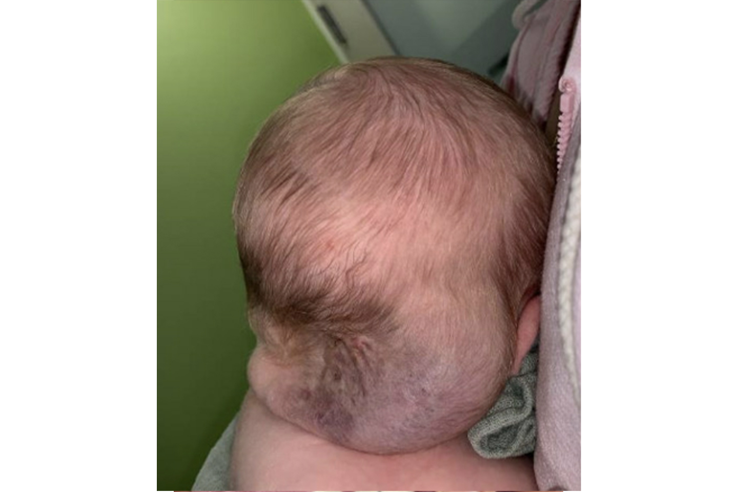
A female infant presented with a recurrently draining and bleeding lymphovenous malformation on her scalp, combined with a calvarial defect. Source: NYU Langone Health.
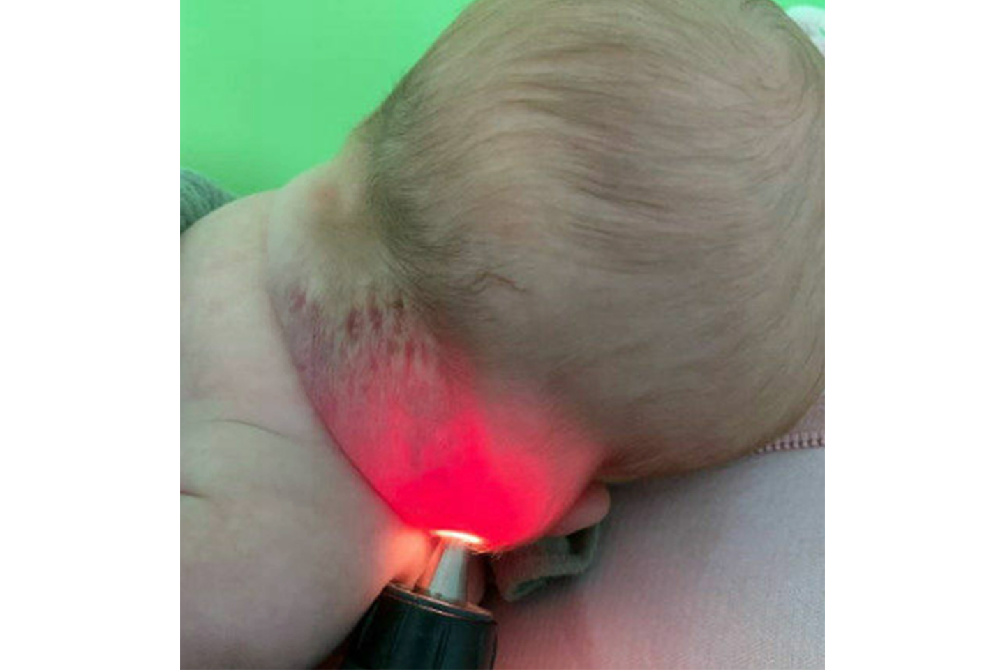
The infant’s compressible mass showed serosanguinous drainage transillumination, which indicated a buildup of bloody proteinaceous fluid. Source: NYU Langone Health.
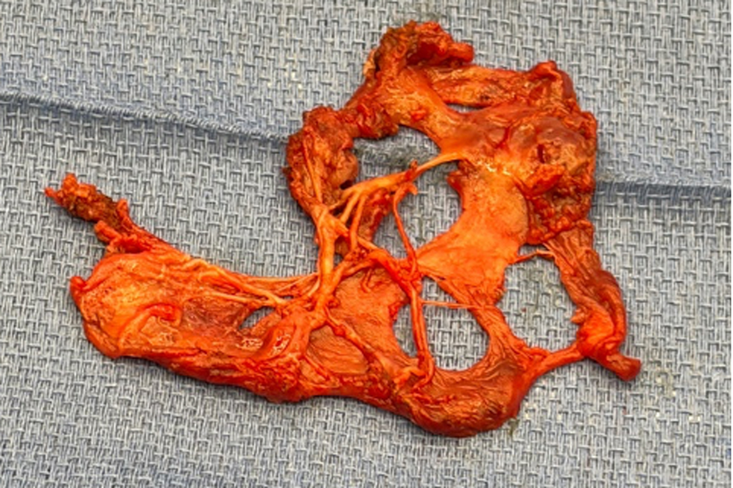
Surgeons removed 90 percent of the cyst wall from the patient’s lymphovenous mass, and pathology tests confirmed that the mass was benign. Source: NYU Langone Health.
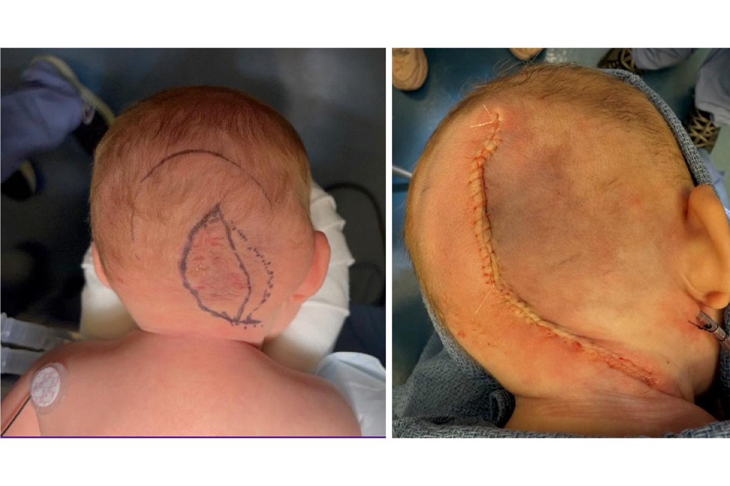
Surgeons also removed involved scalp skin in order to reduce the risk of recurrence (L), and closed the incision with an advancement flap procedure (R). Source: NYU Langone Health.
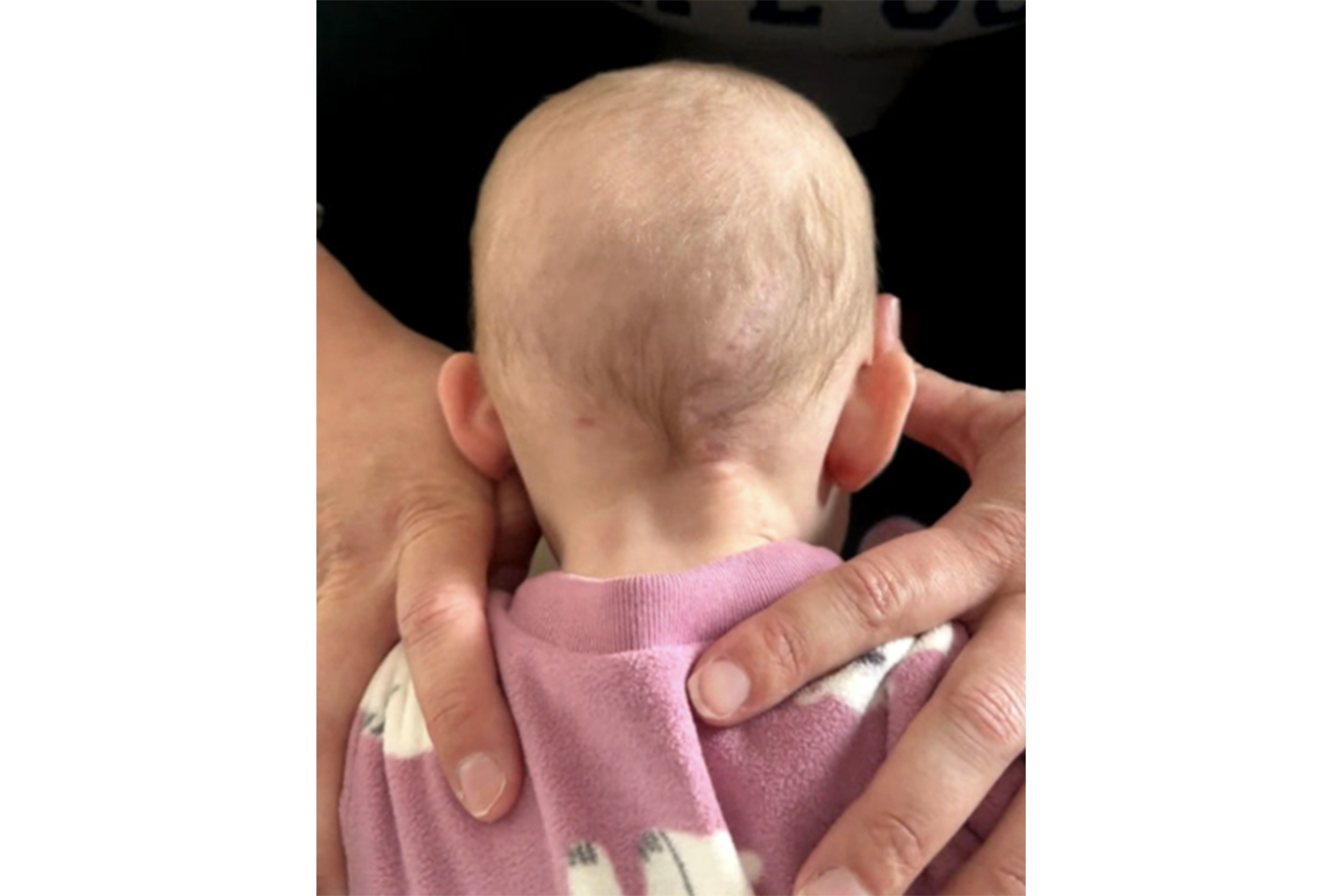
At a follow-up appointment, the patient’s scar had healed well. Source: NYU Langone Health.