
Patellofemoral instability is a relatively common condition among the young active population and has a multifactorial etiology. Both soft tissue constraints, such as the medial patellofemoral ligament (MPFL), and bony geometry confer patellar stability. The relationship between the tibial tubercle and the trochlear groove, as well as the geometry of the trochlea itself, are critical considerations. We now appreciate that a shallow or dysplastic trochlea plays a significant role in recurrent patellofemoral instability.
Conservative options, such as bracing and physical therapy, are often effective initial treatments. However, in patients with recurrent instability, surgical intervention is indicated. In recent years, groove-deepening trochleoplasty has gained further interest as an adjunct to more classically described soft tissue surgical techniques. Although technically demanding, when combined with soft tissue reconstructions, trochleoplasty has been shown to be a powerful tool to correct bony tracking abnormalities.
Here we present a case of a 26-year-old female with a history of recurrent patellar instability and underlying trochlear dysplasia, successfully treated with a combined trochleoplasty and soft tissue Medial Quadriceps Tendon Femoral Ligament (MQTFL) reconstruction.
Case Highlights:
- The patient presented with a history of recurrent left knee patellar dislocations and inability to return to sports.
- Preoperative imaging demonstrated severe trochlear dysplasia (Dejour B/D, shallow/convex trochlea, positive spur and crossing signs) and significant lateral patellar tilt.
- The patient underwent a thin flap groove-deepening trochleoplasty fixed with knotless anchors, combined with MQTFL reconstruction and lateral lengthening.
- The postoperative course was complicated by arthrofibrosis requiring manipulation under anesthesia at 4 weeks, after which she regained full range of motion.
- At approximately 2.5 years postoperatively, she is doing well, biking and hiking with minimal discomfort, and reports high satisfaction with her functional recovery.
Case Presentation
The patient is a 26-year-old female who presented with left knee pain and instability. She reported a history of two prior dislocations, the first occurring 2 years ago, followed by multiple subsequent subluxations and dislocations. At the time of presentation, she was unable to return to sport and exercise due to pain and apprehension. Her past medical and surgical history was unremarkable.
On physical examination, she had a mild effusion with neutral mechanical alignment bilaterally. She demonstrated a lateral resting position of her patella and tenderness to palpation at the medial patellar facet. Her range of motion was full extension to 110 degrees of flexion. Clinically, she had a +3 lateral translation with a glide test without a firm endpoint, a positive patellar apprehension test, a positive J sign, and positive lateral patellar tilt.
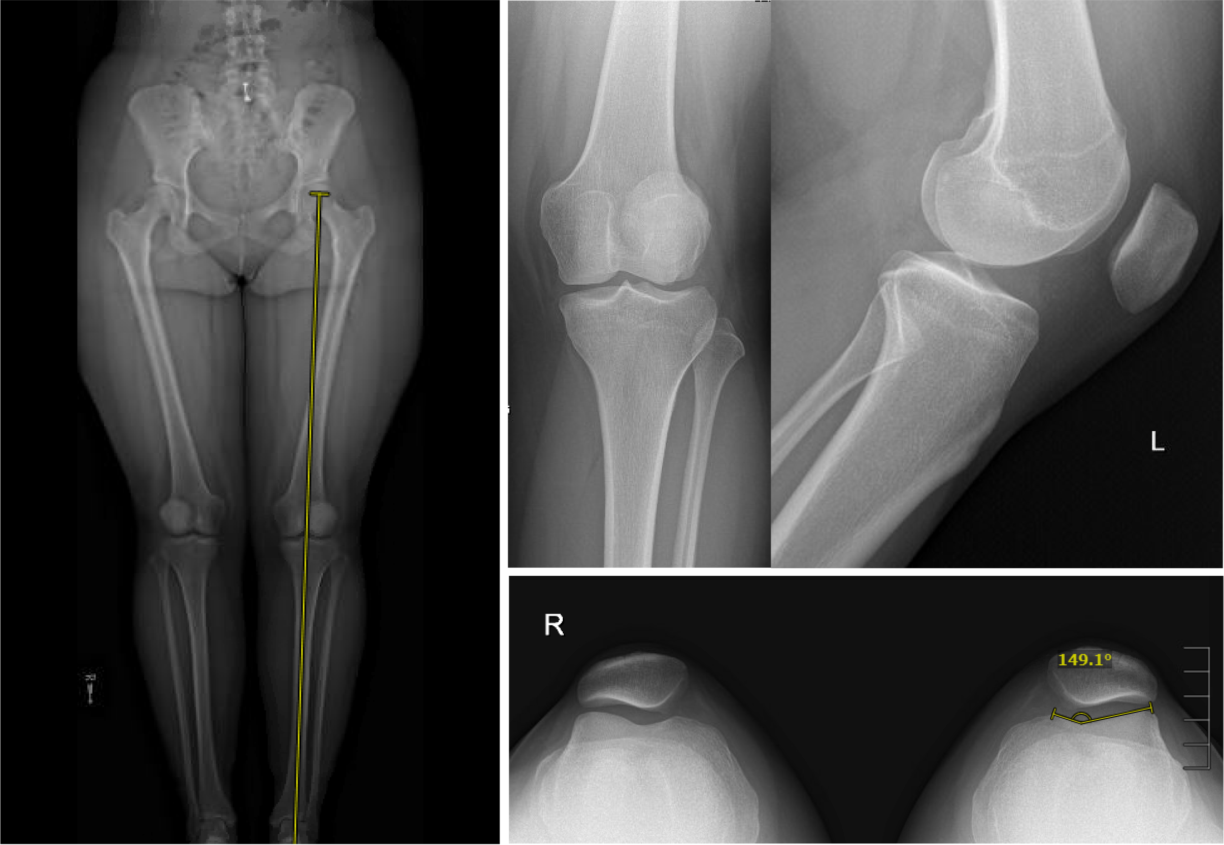
Preoperative imaging confirmed neutral mechanical alignment on full-length standing films. Radiographs demonstrated a Caton-Deschamps ratio of 1.18, a positive supratrochlear spur sign, a positive crossing sign, and a shallow trochlea (Figure 1).
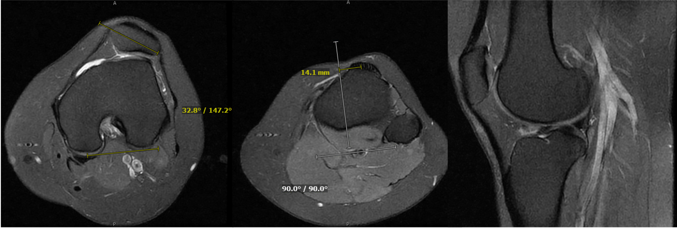
Advanced imaging (MRI) revealed a flattened/convex trochlea, lateral patellar tilt of 32.8 degrees, and a TT-TG distance of 14.1 mm (Figure 2). Given her age, inability to perform activities, and extremely dysplastic trochlea, she was indicated for surgical management including a trochleoplasty and soft tissue reconstruction.
Operative Management
After induction of anesthesia, an examination under anesthesia was performed, demonstrating a laterally dislocating patella and a positive J sign (Video 1).
Diagnostic arthroscopy revealed a dysplastic trochlea, undersurface fraying of the patella, and confirmed lateral subluxation upon stress examination.
A standard lateral parapatellar approach was performed, isolating the superficial and deep layers to allow for a lateral lengthening at the conclusion of the procedure. An arthrotomy was made along the lateral aspect of the trochlea and patella.
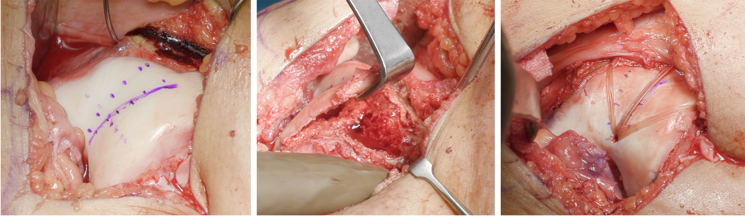
The native and desired center of the trochlea were identified and a small osteotome was methodically used to elevate a thin osteochondral flap circumferentially, taking care not to violate the cartilage surface (Video 2).
Once the subchondral surface was freed, a burr was used to excavate the undersurface of the trochlea and subchondral bone, creating space for plastic deformation of the native cartilage surface (Video 3). Superior lateral trochlear spurs were sharply debrided. The osteochondral flap was then depressed to deepen the groove into the recessed bone.
Fixation was achieved using four preloaded knotless anchors with #2 absorbable suture placed along the superior pole of the trochlea and in the intercondylar notch (Video 4, Figure 3). The sutures were carefully tensioned to secure the new trochlea without causing cartilage necrosis.
Attention was then turned to the medial soft tissue reconstruction utilizing an MQTFL technique (Video 5). A small rent was made in the superior medial aspect of the vastus medialis obliquus (VMO) at Tanaka’s point. Schottle’s point was identified on the femur fluoroscopically, a guide wire passed, and over-reamed to the lateral cortex. A soft tissue allograft was passed into the femur and secured with a single interference screw. The graft was then passed through the VMO rent.
Prior to securing the medial graft, the lateral arthrotomy was closed with a lateral lengthening to prevent over-tensioning and over-medializing the patella. The MQTFL graft was then appropriately tensioned and secured to the quadriceps tendon using multiple FiberWire sutures. Final range of motion and manual stress testing demonstrated a secure patella gliding centrally within the new, deepened trochlea without the ability for lateral subluxation.
Postoperative Course
Immediately postoperatively, the patient was allowed partial weight-bearing with the knee locked in full extension. Between 2 to 6 weeks, she was transitioned to weight-bearing as tolerated, still locked in extension, and began progressing range of motion in physical therapy. The brace was discontinued between 6 and 12 weeks once quadriceps function returned.
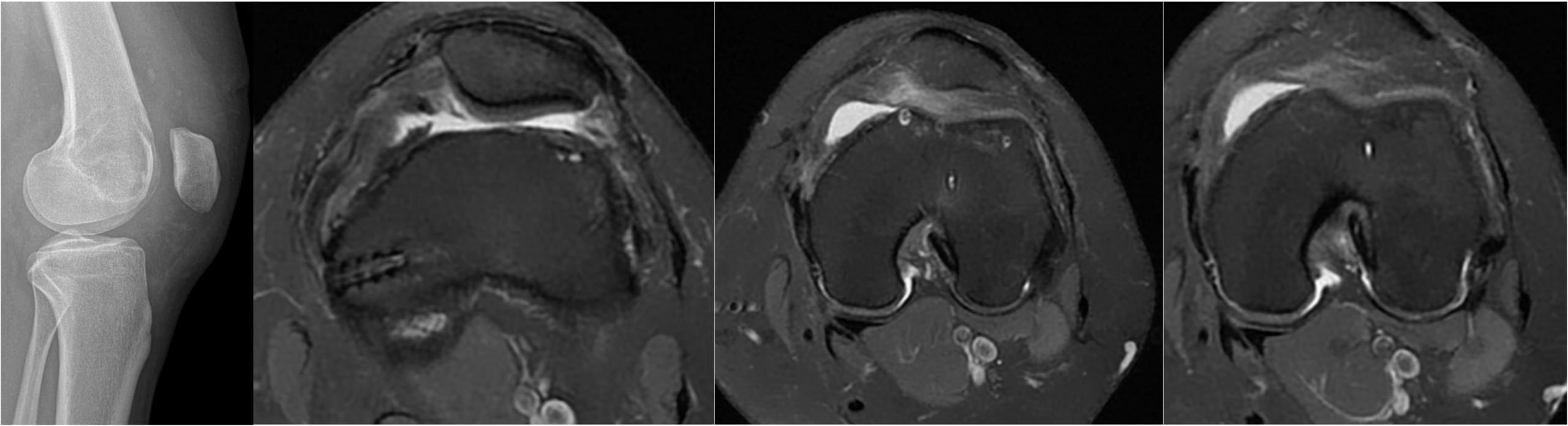
Her initial follow up was complicated by reduced range of motion and arthrofibrosis, necessitating a manipulation under anesthesia (MUA) at 4 weeks postoperatively. Following the MUA, she successfully regained her motion. At 6 months, she had full, painless range of motion (0 to 130 degrees) and had resumed full activities. Postoperative imaging, including a 4-week lateral radiograph and a 6-month MRI, showed good alignment, maintenance of the deepened trochlear groove, and no evidence of patellofemoral osteoarthritis (Figure 4).
At 1 year postoperatively, clinical examination demonstrated full painless range of motion, no maltracking, and a pain-free non-antalgic gait (Video 6). The patient described no feelings of patellofemoral instability.
The patient was most recently evaluated at approximately 2.5 years post-op. She continues to have good results. She is able to do everything she wants to do, including biking with no discomfort. She denies any other symptoms and states her knee is functioning very well overall.
Discussion
Management of recurrent patellar instability in the setting of severe trochlear dysplasia remains challenging. While isolated soft tissue procedures like MPFL reconstruction are effective for many patients, those with significant bony abnormalities—such as a convex or flattened trochlea—often require bony correction to restore stability and normal patellofemoral kinematics.
Trochleoplasty relies on reshaping the dysplastic distal femur to create a stable groove for patellar tracking. Literature shows favorable outcomes, with recent systematic reviews demonstrating low recurrence rates, particularly when trochleoplasty is combined with MPFL reconstruction. A meta-analysis of over 1,000 trochleoplasties showed positive overall outcomes, though a subset of patients developed moderate patellofemoral osteoarthritis or required recurrent surgery, underscoring the procedure’s technical demands and risks. Isolated trochleoplasty has been associated with slightly higher residual instability compared to combined soft tissue and bony reconstruction procedures.
This case highlights several key surgical pearls. First, marking the native and desired center of the trochlea is essential to properly align the new groove. Second, preservation of the lateral femoral condyle height is critical. Third, extreme care must be taken to thin the undersurface without penetrating the articular cartilage, and fixation sutures must not be over-tensioned, which could precipitate cartilage necrosis.
In summary, groove-deepening trochleoplasty combined with medial soft tissue reconstruction and lateral lengthening is an effective, albeit technically demanding, surgical option for young, active patients with recurrent instability and high-grade trochlear dysplasia. With careful technique and appropriate rehabilitation, patients can achieve excellent functional outcomes and return to their desired activities.