Virtual reality (VR) is becoming an important tool in the field of neurosurgery, with applications to medical training and education, as well as in surgical planning and guidance.
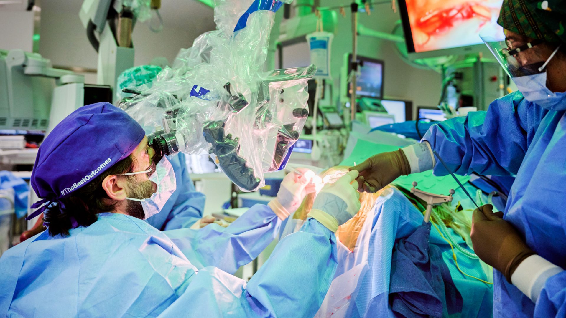
At NYU Langone Health, a team of neurosurgeons led by Erez Nossek, MD, director of the Cranial Bypass Program, is piloting VR technology for preoperative planning of superficial temporal artery (STA)–to–middle cerebral artery (MCA) bypass.
“VR is enabling surgeons to tailor approaches to a patient’s specific neurovascular anatomy using interactive 3D models that can be viewed in high resolution.”
Erez Nossek, MD
“VR is enabling surgeons to tailor approaches to a patient’s specific neurovascular anatomy using interactive 3D models that can be viewed in high resolution,” Dr. Nossek says. “These models have been used in STA-MCA bypass planning to achieve the minimum necessary craniotomy size for a precise bypass localization.”
Precise Planning
The complexity and intricate nature of STA-MCA bypass warrants precise preoperative planning to identify the appropriate STA donor and MCA recipient, Dr. Nossek explains.
“We have to find the optimal location and size of the craniotomy to ensure a high patency rate, as well as low rates of surgical morbidity and mortality.”
In contrast to conventional imaging, the VR technology renders 360-degree models of patient-specific anatomy from volumetric imaging, providing enhanced visualization of the spatial relationship within the targeted surgical anatomy and allowing the surgeon to view the patient’s anatomy in high resolution from any vantage point.
Efficient Procedures
Dr. Nossek and his team recently reported their early experience using the VR-based technology for preoperative planning of STA-MCA bypass. Caleb Rutledge, MD, an assistant professor of neurosurgery and director of cranial and vascular neurosurgery at NYU Langone Hospital—Brooklyn, is a co-author on the study.
From August 2020 to February 2022, 30 patients undergoing STA-MCA bypass were enrolled into two groups. In one group, a VR-based surgical planning platform was used to preoperatively plan the procedure. In the control group, the surgeon used conventional preoperative planning techniques.
“The VR platform facilitated smaller and more precise craniotomies, allowing for an easier and more efficient anastomosis.”
“The VR platform enabled the selection of the ideal vessel candidates for anastomosis and localization of the center of the craniotomy with respect to the anastomosis site,” Dr. Nossek says. “The platform facilitated smaller and more precise craniotomies, allowing for an easier and more efficient anastomosis.”
Blueprint for the Future
While these data are preliminary, Dr. Nossek and Dr. Rutledge are excited about the future of the technology and its many potential benefits, including the ability to be used as a tool for trainees to learn the surgical techniques.
“For recipient vessel selection, the VR system facilitated the selection of the best potential donor and recipient, which might improve patency in larger-scale studies,” Dr. Nossek says. “From an educational perspective, it may be a useful tool for trainees to understand the vascular anatomy and relationships between potential donors and potential recipients.”


