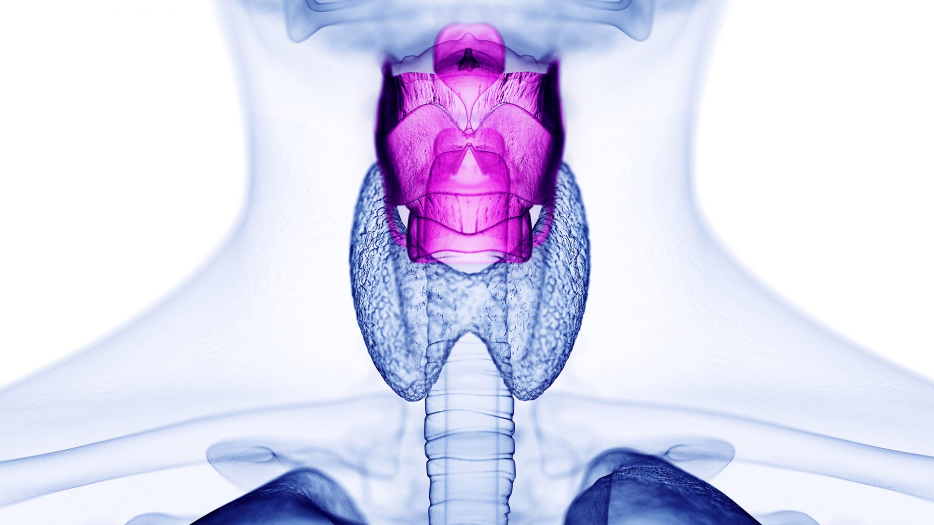
Laryngeal diseases are commonly divided into two mutually-exclusive diagnostic categories: mucosal injury or neuromuscular/functional disorders. Yet this distinction does not account for potential interactions between tissue types, as occurs in other organ systems. Until recently, no such relationship had been described in the vocal folds.
Recently, NYU Langone Health researchers published the first-ever study of interactions between cells from the vocal fold mucosal and underlying muscle in Annals of Otology, Rhinology and Laryngology. Led by Ryan C. Branski, PhD, the Howard A. Rusk Associate Professor of Rehabilitation Research, the team included Ryosuke Nakamura, PhD, an associate research scientist in the Department of Rehabilitation, and Aaron M. Johnson, PhD, associate professor of rehabilitation medicine and otolaryngology-head and neck surgery. The researchers used tissue from rats, culturing mucosal fibroblasts and thyroarytenoid myoblasts in vitro.
“The etiology of vocal fold disorders is diverse, and the efficacy of current interventions is variable. We need to gain a deeper understanding of basic disease processes to improve outcomes for patients with these conditions.”
Ryan C. Branski, PhD
“The etiology of vocal fold disorders is diverse, and the efficacy of current interventions is variable,” Dr. Branski explains. “We need to gain a deeper understanding of basic disease processes to improve outcomes for patients with these conditions.”
Seeking Clues from Lower Airway Disorders
The team investigated whether a phenomenon known to occur in the lower airway might apply to the vocal folds as well: interactions between mucosal and muscular tissue. In asthma, for example, increased contractile properties of airway smooth muscle are considered a hallmark of the disease, together with mucosal inflammation.
This combination of pathologies appears to be related to altered expression of contractile proteins—particularly, myosin light-chain phosphorylation—within airway smooth muscle. Downstream, overexpression of fast myosin heavy chain isoforms in airway smooth muscle enhances intracellular calcium signaling, Dr. Branski explains. Overexpression of calcium-sensing receptor, in turn, further exacerbates the abnormal cascade of mucosal-muscle signaling in asthma.
A Surprising Reciprocity
Dr. Branski and his colleagues hypothesized that injury to the vocal folds elicits similarly complex interactions between the vocal fold lamina propria and the underlying muscle. “As a first step to test this idea, we sought to investigate the effects of vocal fold fibroblasts on myoblasts,” he says. “Interestingly, however, our data show significant bidirectional effects between cell types.”
“These findings suggest that laryngeal muscle health may contribute to mucosal well-being. Conversely, muscle dysfunction could potentially have a deleterious effect on the vocal fold mucosa.”
The researchers analyzed fibroblast proliferation and migration in the presence of myoblast-conditioned growth media, using TGF-beta and other substances to induce the myofibroblastic phenotype. After 24 hours, they analyzed phenotypic shifts and cellular differentiation.
Generally, the myoblasts favorably impacted fibroblasts (for example, decreasing Acta2 mRNA expression), while the effects of fibroblasts on myoblasts were unfavorable (for example, decreasing myosin heavy chain expression).
“These findings suggest that laryngeal muscle health may contribute to mucosal well-being,” Dr. Branski observes. “Conversely, muscle dysfunction could potentially have a deleterious effect on the vocal fold mucosa.”
The potential implications of these interactions warrant further investigation, he stresses. Ultimately, such research could lead to better treatment strategies—whether pharmacological, surgical, or behavioral—for a wide range of vocal fold disorders.