
Urology
Surgical Innovation
New SP Technique Advances Right-Sided Donor Nephrectomy
Surgeons develop a single-port, retroperitoneal approach for kidney donation that supports rapid recovery without compromising outcomes.


NYU Langone Health: A Leader in Urology
Ranked #2
in Urology
Top 5
in NIH funding (Source: Blue Ridge)
Pioneers in MRI
to evaluate prostate cancer
Complex Case Spotlight
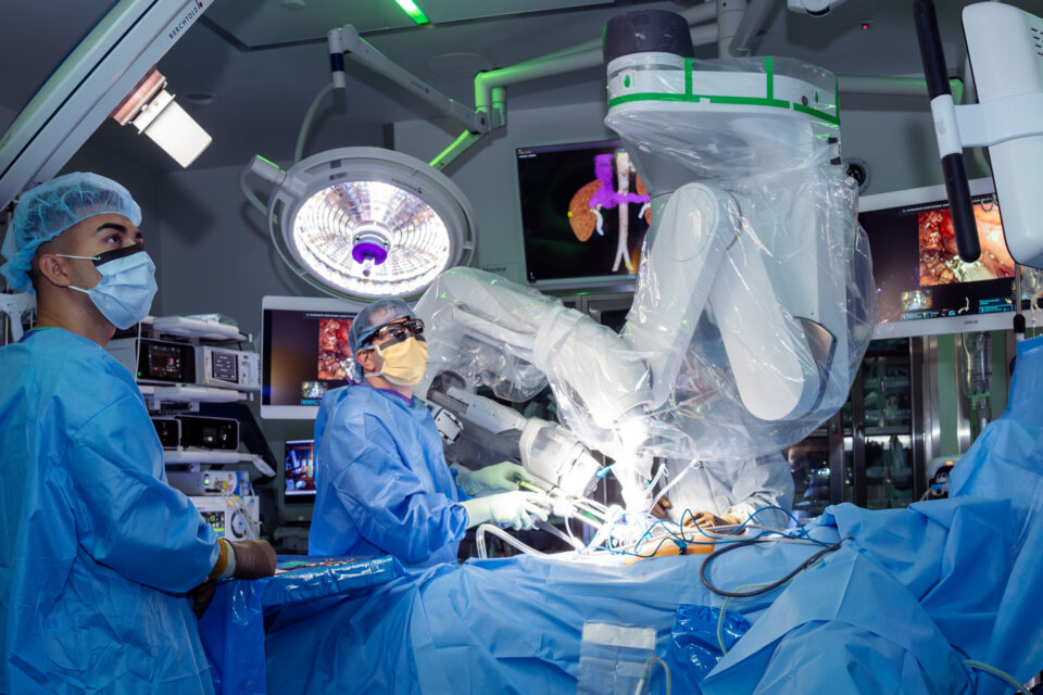
Precision Robotic Prostatectomy: Maximizing Function While Maintaining Oncologic Control
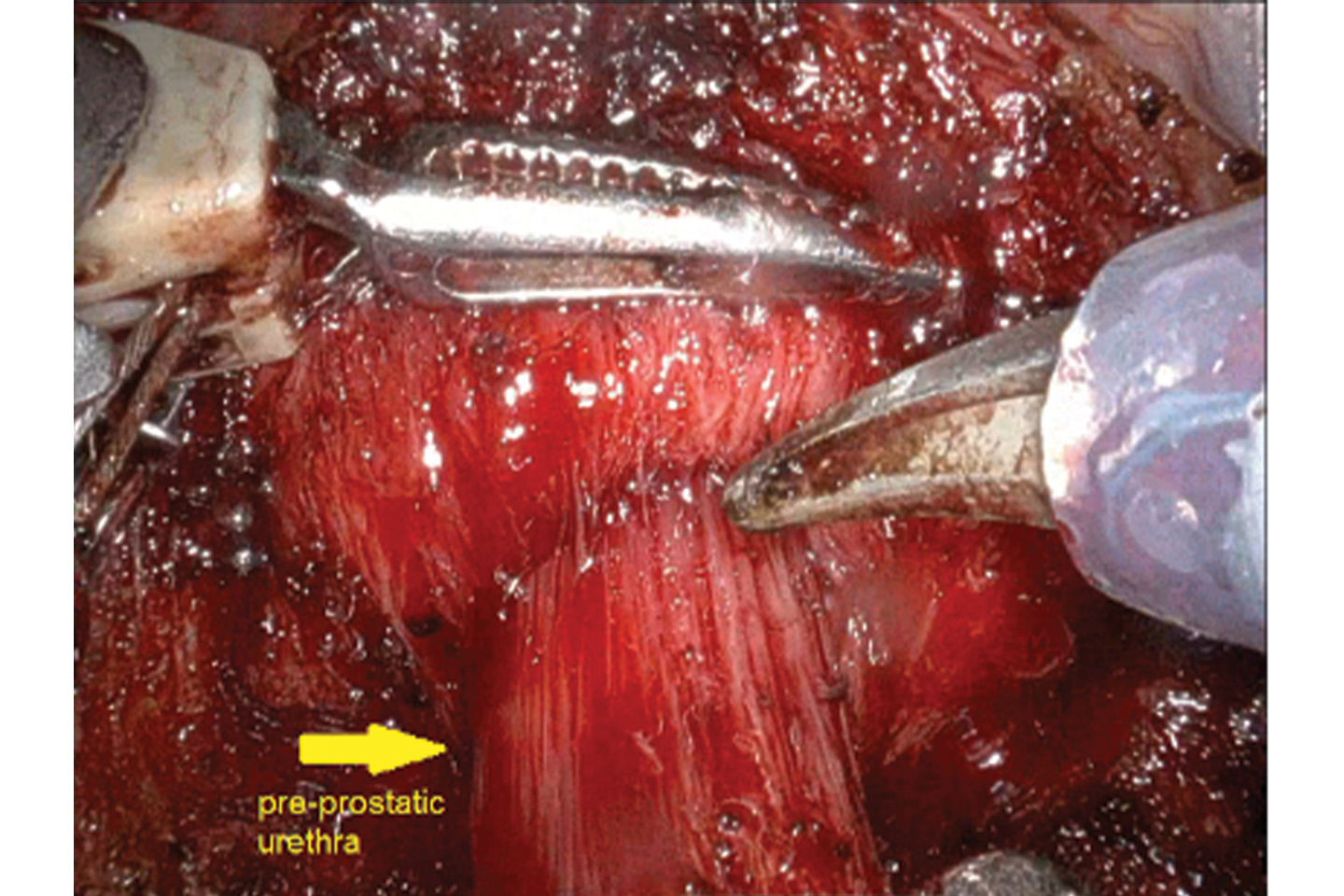
Figure 3A. The pre-prostatic urethra is identified between the bladder neck and prostate. The urethra is carefully dissected circumferentially to delineate its full length and maintain maximal preservation prior to transection. Source: NYU Langone Health.
1 of 5
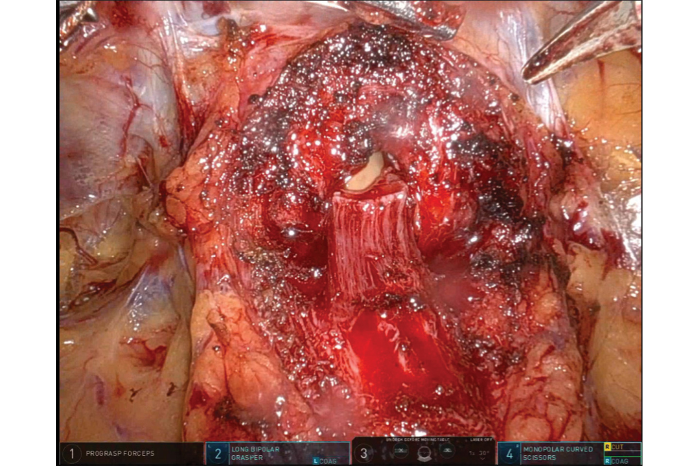
Figure 3B. Precise incision of the pre-prostatic urethra. The urethra is sharply divided under direct vision to maximize urethral length. Source: NYU Langone Health.
2 of 5
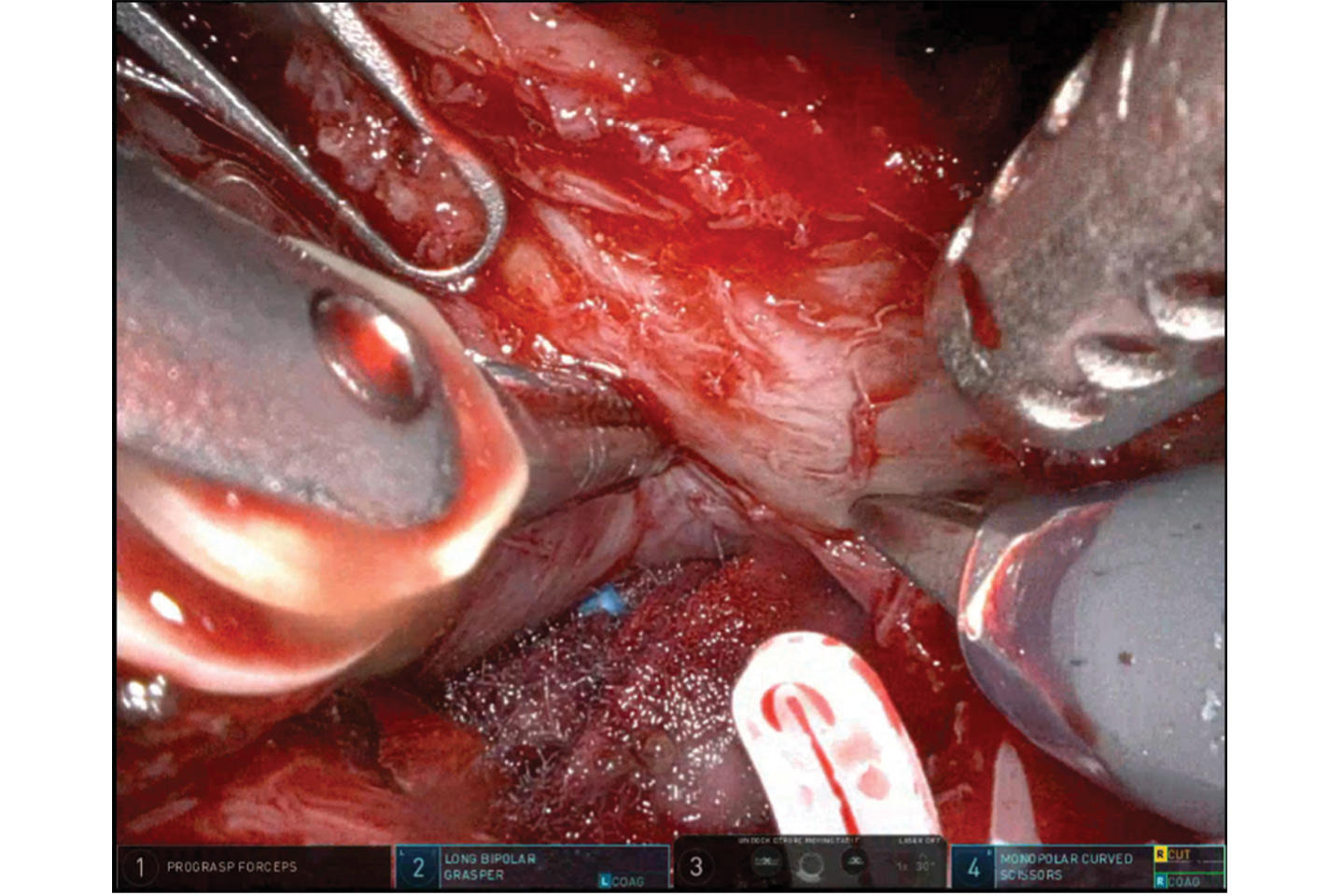
Figure 4. Right-sided intra-fascial nerve-sparing dissection performed with the neurovascular bundle clearly preserved in close proximity to the prostatic capsule. The dissection plane was developed within the prostatic fascia, demonstrating meticulous preservation of periprostatic tissue integrity. Source: NYU Langone Health.
3 of 5
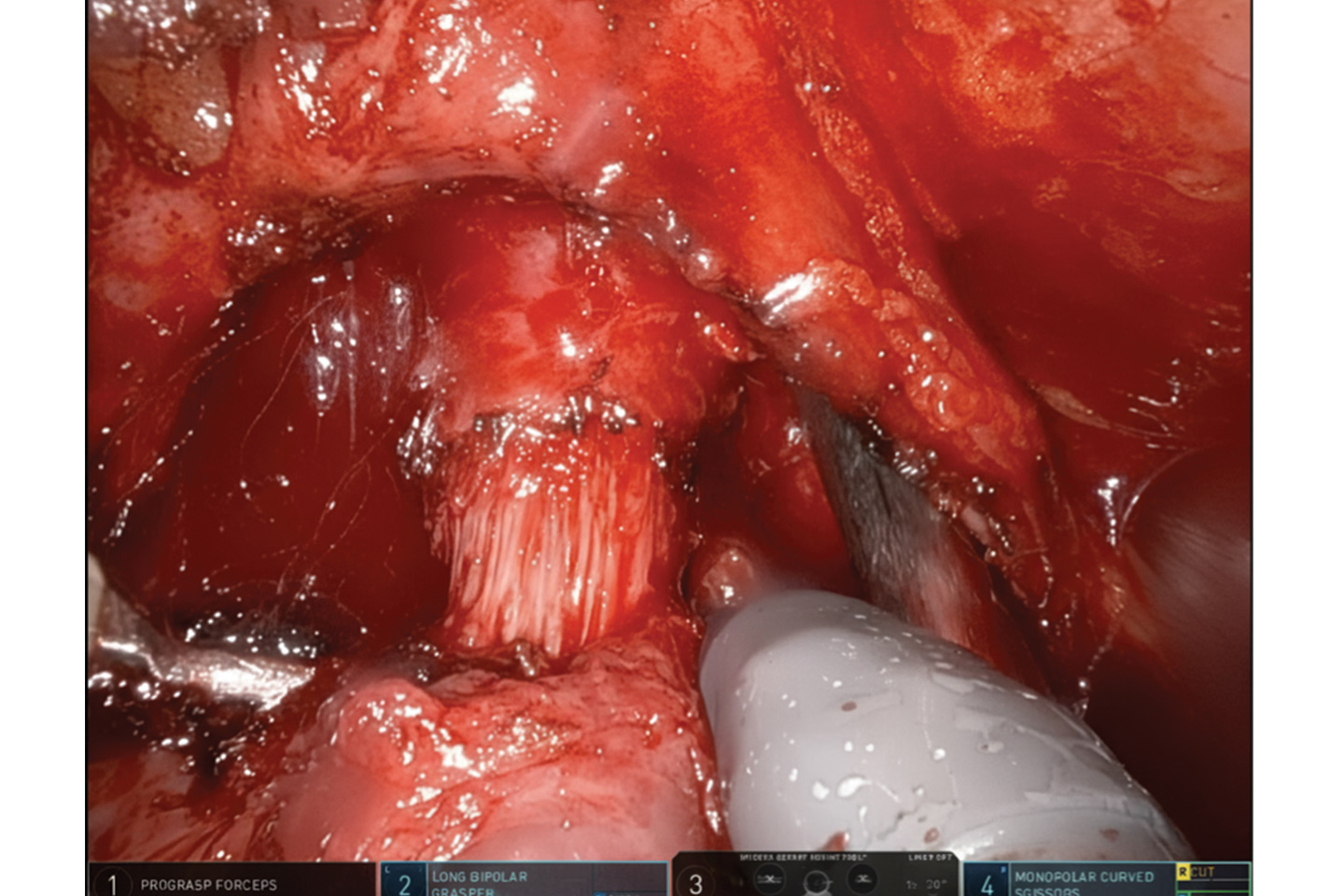
Figure 5. Preservation of the apical prostatic urethra. The distal prostatic urethra is carefully dissected free from the prostatic apex. Source: NYU Langone Health.
4 of 5
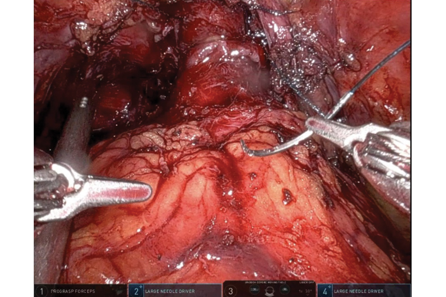
Figure 6. Maximal urethral preservation technique was employed, sparing an additional 2 cm of the pre-prostatic urethra and the apical prostatic urethra, allowing reconstruction of a tension-free anastomosis resembling a urethroplasty. Source: NYU Langone Health.
5 of 5

The Best Experts and Latest Breakthroughs
Select your specialty to receive updates on our pioneering research, innovations, expert perspectives, case studies, practice-changing medicine, and more.