Correction of Dissociated Antibiotic-Loaded Hip Spacer
A 65-year-old woman who had undergone a total hip arthroplasty for avascular necrosis in her left hip subsequently contracted a bacterial infection that required several revision surgeries at NYU Langone Health to implant an antibiotic-loaded temporary hip spacer and treat her trochanteric fracture and instability.
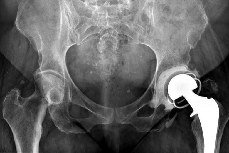
Twenty months after revision, the patient presented to the emergency department after experiencing discomfort and noticing a significant change in her leg length for three to four days. Pelvic X-rays revealed a severe dislocation of the hip spacer with near-complete extraction of the cemented implant from the left femur.
Prior history of trochanteric fracture added to the patient’s hip instability. Orthopedic surgeon Morteza Meftah, MD, worked with colleagues to confirm the absence of infection and performed a same-day revision surgery in which he converted the spacer to a final implant secured with screws and a constrained liner.
“The challenges of the case were ruling out infection, addressing a four-day-old dislocation in a non-compliant patient with pressure on her sciatic nerve, and managing the loss of her trochanter and the resulting instability from a lack of attached abductor muscles,” Dr. Meftah says. “We also had to meticulously remove all of the cement from her femur and acetabulum and take into account her stiff spine to avoid implant impingement while getting her leg length equal.”
“We managed to rule out infection, get the appropriate implants, and clear her right away for surgery knowing that she had been dislocated for several days.”
Morteza Meftah, MD
Computer navigation helped the surgical team ensure an anatomy-appropriate correction of the patient’s leg length and proper implant positioning.
“It’s teamwork, and that includes infectious disease, anesthesia, and the entire perioperative surgical teams,” Dr. Meftah says. “We managed to take care of this patient very rapidly in a very safe manner to rule out infection, get the appropriate implants, and clear her right away for surgery knowing that she had been dislocated for several days.”
Two weeks post-surgery, the patient was walking well with a cane and pleased with her progress.
X-Ray of an Antibiotic-Loaded Hip Spacer
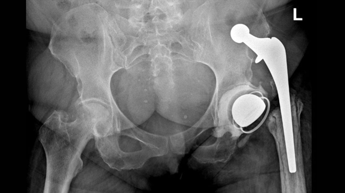
One year after a total hip arthroplasty for avascular necrosis in her left hip, the patient sustained a periprosthetic dislocation with trochanteric fracture and chronic bacterial infection. She underwent revision surgery to implant an antibiotic-loaded hip spacer with a constrained liner. The loss of her left trochanter is visible in this X-ray.
Hip Implant Dislocation and Dissociation
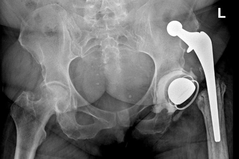
Twenty months after the surgical insertion of the temporary hip spacer, the patient suffered a severe dislocation with a near-complete extraction of the cemented implant from the left femur.
A Same-Day Correction
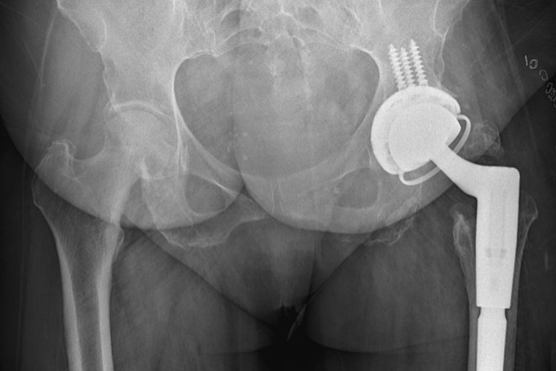
A same-day revision surgery converted the temporary spacer to a final implant secured with screws and a constrained liner.