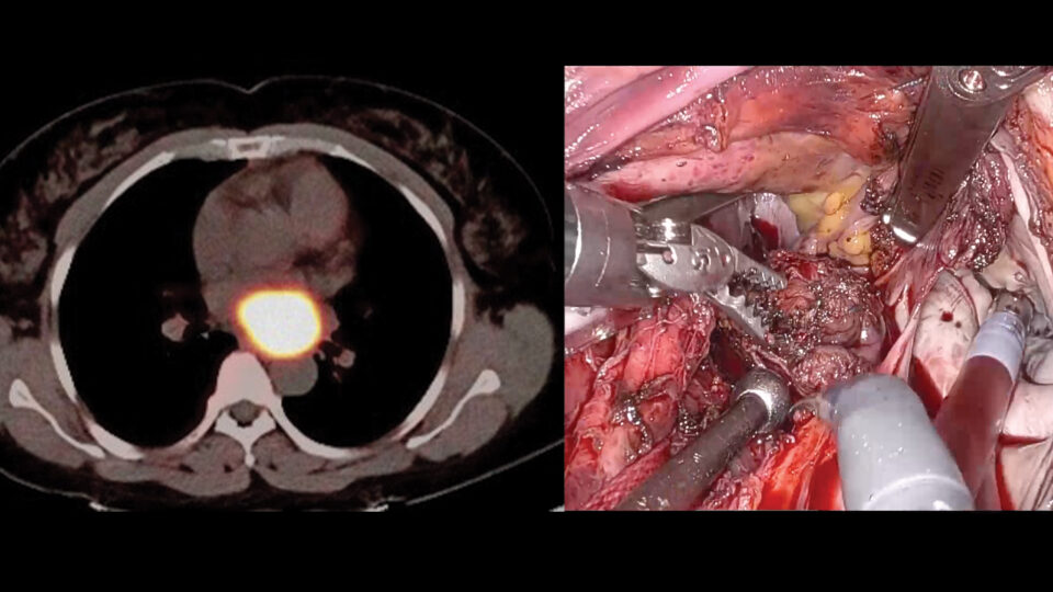
Diagnosing suspicious peripheral pulmonary lesions seen on CT scan has often required bronchoscopy and an additional thoracic surgery to retrieve the tissue needed for biopsy.
Interventional pulmonologists Jamie L. Bessich, MD, and Vivek Murthy, MD, are using robotic bronchoscopy to improve minimally invasive diagnosis of lung nodules, reducing the need for more invasive procedures.
NYU Langone Health was involved in testing the robotic equipment and was one of the first clinical sites in the U.S. to use the technology.
“The paradigm has shifted in interventional pulmonology,” says Dr. Bessich. “With CT scans emitting less radiation than before and lung cancer screening widely available, the incidence of finding lung lesions is a lot higher. As a result, we’re seeing more patients needing biopsies and need a really accurate way to get to these lesions.”
Increasing Diagnostic Yield
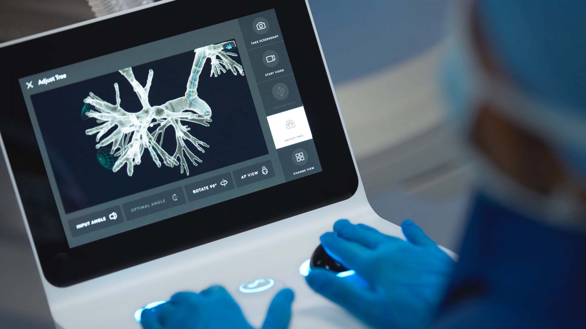
Robotic bronchoscopy builds on technical and procedural advances for minimally invasive diagnosis of lung nodules developed since the introduction of endobronchial ultrasound in the 1990s. It allows for fine control of bronchoscopic navigation by replacing hand-directed movements with electronic control mechanisms.
An ultrathin, ultra-flexible catheter allows navigation far into the periphery of the lung, enabling the precision needed for biopsy.
“The robotic software generates a high-resolution map of a patient’s lungs and helps us direct the minimally invasive tools to target and biopsy lung nodules without making any cuts in the skin.”
Vivek Murthy, MD
“The robotic software generates a high-resolution map of a patient’s lungs and helps us direct the minimally invasive tools to target and biopsy lung nodules without making any cuts in the skin,” Dr. Murthy explains. Once a target is identified, the three-dimensional image displays airway trees and automatically generates a path and anatomic borders to maximize safety.
Dr. Bessich says the robot has a much better yield than all prior navigational software in finding, accessing and biopsying suspicious nodules.
“It used to be that if a patient needed biopsy of a lymph node, we would do that procedure separately, and the nodule would often be accessed via CT-guided biopsy, coming from the outside of the chest. Now, with the improved yield from robotic bronchoscopy, we’re doing them both together and potentially eliminating the need for a second procedure.”
Enhancing Cancer Diagnosis
The interventional pulmonology team is working with NYU Langone neurology colleague Daniel Orringer, MD, to combine robotic bronchoscopy with Raman spectroscopy in evaluating nodules they suspect are cancerous—without destroying any more tissue than necessary.
Raman spectroscopy is a light scattering technique that stimulates cells and tissue and captures a high-fidelity graphic image, which is colored to resemble a hematoxylin and eosin (H&E) pathologic stain.
“Because of the quality, SRH (stimulated Raman histology) allows pathologists to look for signature cancer cells. They feel more comfortable because it looks like the familiar H&E stain,” Dr. Bessich says. “When we do forceps biopsies, we need to take eight to 10 samples; we are giving them one or two samples from SRH to analyze.”
Currently, pathologists aren’t reviewing the images in real time; the pulmonologists are saving the images and sending to them. The hope moving forward is that with robotic bronchoscopy, they will eventually have real time, on-site confirmation that peripheral pulmonary lesions need ablation or resection.
Future Directions
As they employ robotic bronchoscopy for more and more cases, Drs. Bessich and Murthy are using the artificial intelligence features of the equipment, teaching it to distinguish cancerous lesions from noncancerous. “The more airways we go into and the more pictures we get, the more accurate it gets,” Dr. Bessich says.
Their goal is to identify the minimum volume of tissue needed for any molecular or genetic testing an oncologist wants completed. Their eventual hope is to be able to apply therapeutics using the robotic bronchoscope.
“The beauty of doing everything via bronchoscopy is that we spare the patient multiple procedures, while at the same time getting better outcomes.”
Jamie L. Bessich, MD
“Robotic bronchoscopy has revolutionized our approach,” Dr. Bessich says. “The beauty of doing everything via bronchoscopy is that we spare the patient multiple procedures, while at the same time getting better outcomes.”