While technologies for imaging the cornea have significantly improved through advances in specular microscopy, there are still no commercially available tools capable of imaging the entire corneal endothelium, including its peripheral regions.
Gaining a more complete view of the cornea can provide insight into how the outermost edges respond to treatments and infections, potentially translating to improvements in care for those with vision loss secondary to corneal damage or disease.
At NYU Langone Health, a breakthrough imaging device debuted in 2023: Konan Medical’s CellChek® C. The state-of-the-art, wide-field scanning specular microscope is capable of providing near-complete visualization of the cornea, says Kathryn A. Colby, MD, PhD, the Elisabeth J. Cohen, MD, Professor of Ophthalmology in and chair of the Department of Ophthalmology at NYU Grossman School of Medicine. Her team is currently leading studies under IRB approval to test its utility. CellChek C is not yet available for commercial sale in the United States.
“Only a handful of these microscopes exist worldwide.”
Kathryn A. Colby, MD, PhD
“Only a handful of these microscopes exist worldwide,” Dr. Colby says. “We are the first institution in North America to acquire the microscope.”
Visualizing the Entire Corneal Endothelium
Most commercially available specular microscopes are capable of imaging the central and paracentral corneal endothelium, yet they fall short in their ability to image cells in the peripheral endothelium.
According to Dr. Colby, the CellChek C microscope offers the ability to view any portion of the cornea, even the typically hard-to-see periphery, from endothelium to epithelium. The microscope’s slit-scanning technology enables high-resolution and high-contrast imaging of the corneal layers, she adds.
“The images from the CellChek C are breathtaking. Instead of only seeing several hundred central endothelial cells, we can now see tens of thousands of cells throughout the entire cornea.”
“The images from the CellChek C are breathtaking,” says Dr. Colby. “Instead of only seeing several hundred central endothelial cells, we can now see tens of thousands of cells throughout the entire cornea.”
“With its broad imaging capability, the CellChek C is a versatile tool for enhancing our understanding of corneal diseases and the response of the cornea to various surgeries, including Descemet stripping only (DSO) and corneal transplants. Additionally, this device can provide insights into the effect of other eye diseases and surgeries, such as glaucoma or cataract, on the cornea.”
Opportunities for Research
From a research perspective, the microscope offers a plethora of new opportunities, one of which is clinical trials. As the director of the NYU Langone Eye Center, Dr. Colby has pioneered many practice-changing trials for some of the most complex corneal diseases in children and adults.
An ongoing study led by Dr. Colby and her team will compare the CellChek C microscope to the CellChek 20, widely considered the gold standard in specular microscopy.
“We have high hopes that CellChek C will outperform the current standard,” she says. “As we collect more data and explore the microscope’s full potential, we look forward to sharing it with the medical community.”
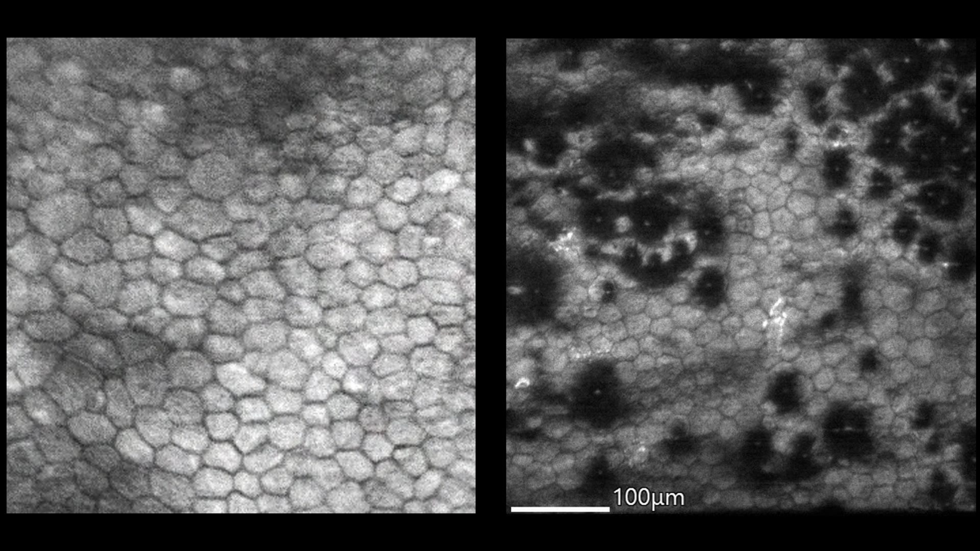
VIDEO: The corneal endothelium in a healthy patient, imaged using the CellChek C.
VIDEO: The peripheral corneal endothelium in a patient with moderate Fuchs dystrophy, imaged using the CellChek C.