Referral Notes:
- CTE still requires post-mortem examination for definitive diagnosis.
- Researchers have identified the first structural features in the brains of living athletes that may indicate elevated CTE risk.
- Structural changes include shallower left superior frontal sulci; longer careers are also associated with wider left occipitotemporal sulci than shorter careers.
- The findings suggest sulcal morphology could help predict CTE risk, though further research is needed.
An international study led by NYU Langone Health reports what may be the first structural differences in the brains of living athletes who may be at higher risk of developing chronic traumatic encephalopathy (CTE).
Despite years of research, clinicians currently must rely on autopsies after death to diagnose CTE. The findings could support the development of tests for early detection.
Study senior author Hector Arciniega, PhD, an assistant professor of rehabilitation medicine at NYU Langone, says the study revealed subtle differences in the sulci of former college and professional football players when compared to brain scans of men who never played contact or collision sports.
“Our study shows what we believe can be the first structural differences that tell apart brains more at risk of developing CTE from the brains of people who are less at risk.”
Hector Arciniega, PhD
“Our study shows what we believe can be the first structural differences that tell apart brains more at risk of developing CTE from the brains of people who are less at risk,” says Dr. Arciniega. “The work also proves that we can apply what we know about the physical changes observed postmortem in the brains of those with confirmed CTE to brain scans of living people at increased risk for it.”
In autopsy analyses, CTE is often marked by shrinking of the brain and the presence of tau protein deposits in sulci near blood vessels.
Shallower and Wider Sulci
The study, published in Brain Communications, is part of a long-term effort to develop tests for early CTE detection.
The researchers analyzed structural MRI data from 169 male former football players and 54 age-matched, unexposed, asymptomatic male controls, all between 45 and 74 years of age. The football cohort included college athletes with at least six years of playing experience and professional players with at least 12 years of experience.
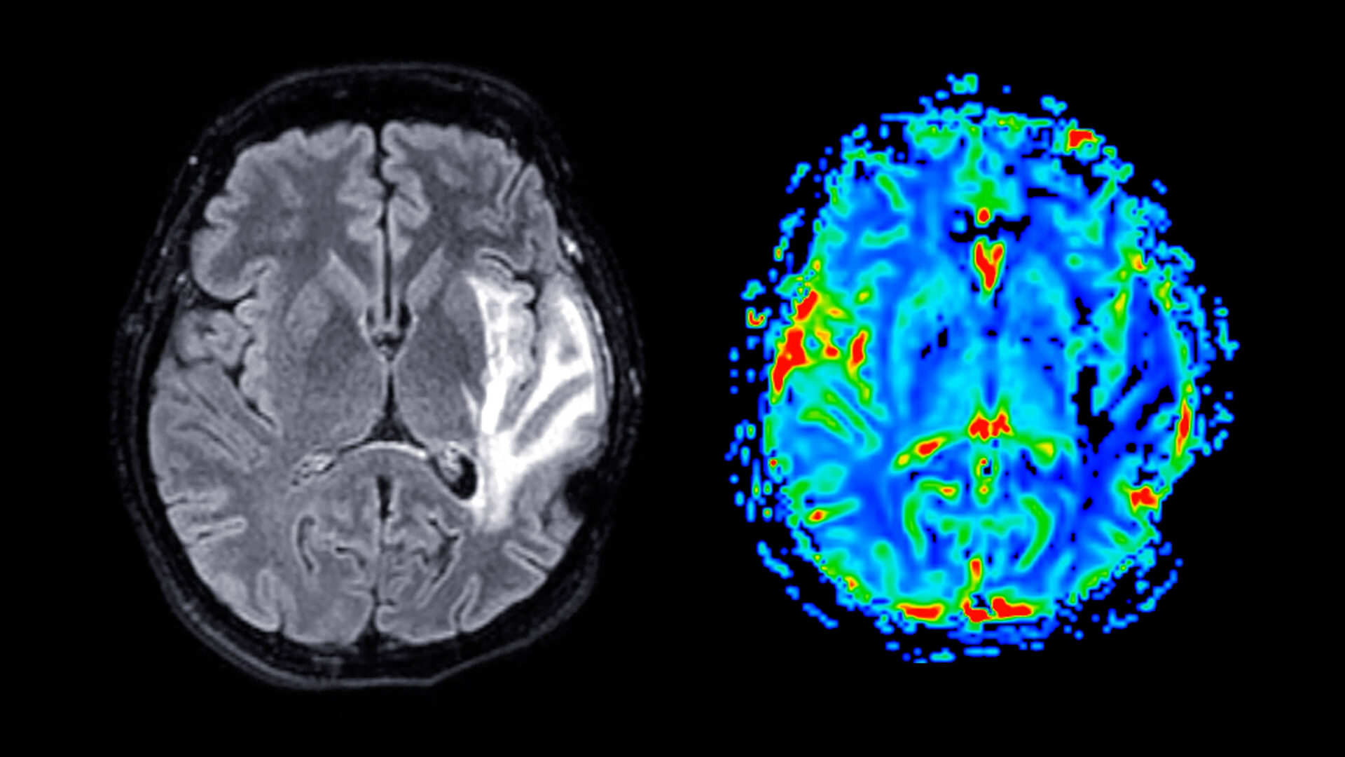
Researchers found that the football players had shallower left superior frontal sulci on average than their non-football counterparts. Left superior frontal sulci are located on a main groove that runs along the top, front, left side of the brain, which is known to be physiologically affected in CTE.
The study also showed that football players with increasing years of playing experience had wider left occipitotemporal sulci than men who started later or had shorter careers. It is unclear why differences were detected only on one side of the brain and not in the sulci on both hemispheres, the researchers say.
While differences in sulci brain structure were shown, no differences were observed regarding comparison of psychological tests for memory and learning, estimates of the number of head hits and injuries, and other brain scan measures of tau.
Regarding a potential pathological mechanism, the authors note that models of brain biomechanics suggest that mechanical strain to the brain concentrates, in part, at the depths of sulci. This localized strain may disrupt axonal integrity and trigger the initiation of tau pathology, which, over time, can drive neuroinflammation, neuronal loss, and cortical atrophy, potentially leading to structural changes of sulcal widening and shallowing, as reported in the study.
Sulcal Morphology as a CTE Biomarker
The sulcal findings could potentially be adopted as biomarkers, Dr. Arciniega says, advancing efforts to develop a diagnostic test so that future therapies can be applied before damage becomes irreversible.
The researchers caution that a clinical diagnostic test remains years away. But they note that if future studies validate their findings, additional biomarkers could be combined, as part of many brain features, into a comprehensive CTE risk assessment.
The team plans to expand their investigation to include more contact and collision sports and test for differences in other parts of the brain to further advance CTE risk prediction.