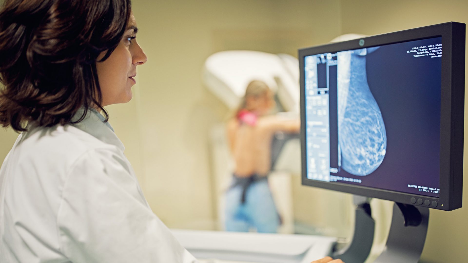
A machine learning model for MRI aids radiologists in detecting breast cancer and may help to reduce unnecessary biopsy, according to a new study published in Science Translational Medicine.
The model is the latest tool from data scientist Krzysztof J. Geras, PhD, and colleagues at NYU Langone Health who are applying artificial intelligence (AI) to improve the accuracy of breast imaging modalities, including mammography and ultrasound. Postdoctoral fellow Jan Witowski, MD, PhD, is first author of the new report.
“Our studies demonstrate how artificial intelligence can help radiologists interpret breast imaging exams to identify only those findings that show real signs of breast cancer and to avoid verification by biopsy in cases that turn out to be benign,” Dr. Geras says.
The suite of models will ultimately be combined into a larger AI system, with the goal of making predictions and learning from multiple imaging modalities simultaneously.
“Our studies demonstrate how artificial intelligence can help radiologists interpret breast imaging exams to identify only those findings that show real signs of breast cancer and to avoid verification by biopsy in cases that turn out to be benign.”
Krzysztof J. Geras, PhD
“We’re hoping to find very subtle correlations among different imaging modalities and learn signal that is too weak for humans to notice,” Dr. Geras says.
AI for Breast MRI
While mammography is largely the relied upon screening exam for breast cancer, MRI has an edge over mammography for its ability to detect small lesions. Yet, a high rate of false-positive findings has limited its use in breast cancer screening, with roughly three biopsies with benign results occurring for every one biopsy with malignant findings.
To improve the specificity of breast MRI, Dr. Geras and colleagues, including imaging specialist Linda Moy, MD, a professor of radiology, used more than 21,000 bilateral dynamic contrast-enhanced breast MRI exams from patients at NYU Langone to build and train a machine learning model. They then evaluated the model on three external datasets from other institutions in the United States and Europe.
“The model is sufficiently robust that it generalizes to other populations. That is often missing in papers that develop AI for different applications,” Dr. Geras says.
Their analysis revealed the AI-based system to be on par with radiologists in detecting breast cancer in MRI exams, and a simulation indicated the model may help to avoid unnecessary biopsies in patients with BI-RADS 4 lesions, for which a “biopsy-all” strategy is currently recommended.
AI for Breast Ultrasound
In a previous study, and one of the largest of its kind, the research team developed an AI model for breast ultrasound, utilizing nearly 5.5 million breast ultrasound images from more than 143,000 patients treated at NYU Langone.
Breast ultrasound rivals both mammography and MRI for its low cost, accessibility, and power to distinguish solid masses from cystic lesions. However, like MRI, difficulty in interpreting images leads to many false-positive findings and unnecessary biopsies.
Not only was the AI model as accurate at generating diagnoses as experienced radiologists, but when used by radiologists to assist in diagnosis, false-positive rates decreased by 37 percent, and the number of requested biopsies dropped by 27 percent.
The study also suggested the system could be used to triage ultrasound exams by automatically dismissing benign cases while escalating high-risk ones to a thorough assessment workflow.
“If our efforts to use machine learning as a triaging tool for ultrasound studies prove successful, ultrasound could become a more effective tool in breast cancer screening…. Its future impact on improving women’s breast health could be profound.”
Linda Moy, MD
“If our efforts to use machine learning as a triaging tool for ultrasound studies prove successful, ultrasound could become a more effective tool in breast cancer screening, especially as an alternative to mammography, and for those with dense breast tissue,” Dr. Moy says. “Its future impact on improving women’s breast health could be profound.”
Further Refining the Models
Dr. Geras cautions that while these initial results are promising, the research teams only looked at past exams in their analyses, and clinical trials of the tools in current patients and real-world conditions are needed before they can be routinely deployed.
He also has plans to refine the AI software to include additional patient information, such as a patient’s added risk from having a family history or genetic mutation tied to breast cancer.


