A 52-year-old female presented to the emergency department at NYU Langone Hospital-Brooklyn complaining of acute-onset middle back pain that had started three days prior. She described the pain as sharp and radiating to her neck, rated the intensity as 8 on a scale of 1 to 10, and stated that it worsened with deep inspiration or other movements.
Three weeks before her visit, the patient had experienced sinus pain, a cough, and a low-grade fever. She tested positive for SARS-CoV-2 and self-quarantined. Her medical history included a diagnosis of scleromyxedema (SM). The condition causes papular and sclerodermoid eruption in the setting of monoclonal gammopathy of undetermined significance. The patient had also been diagnosed with Hashimoto’s disease and asthma, and had been receiving intravenous immunoglobulin (IVIG) for the past seven years. In the ED, however, she did not present with associated symptoms such as fever, chills, shortness of breath, chest pain, abdominal pain, or headache.
“We have learned that SARS-CoV-2 triggers a severe inflammatory response that leads to a cytokine storm and endotheliitis, resulting in arterial and venous thrombosis,” says Yamen Homsi, MD, MPH, who led the rheumatology team involved in the patient’s care. “My initial thought was that our patient could have had an acute pulmonary embolism. However, she lacked the hallmark presenting complaint of chest pain.”
Arriving at an Aortitis Diagnosis
The patient was alert and oriented, and her vitals were normal with the exception of an elevated 154/94 blood pressure reading and heart rate of 90. Her heart, lung, abdomen, and extremities exams were all normal, with no rash, skin tightness, or lymphadenopathy. An initial electrocardiogram and chest X-ray were both unremarkable.
Likewise, the patient had a normal complete blood count and C-reactive protein levels, but an abnormally high erythrocyte sedimentation rate (ESR) of 120 mm per hour, indicating inflammation. The ED team performed a chest CT with IV contrast, which was negative for pulmonary embolism but showed distal aortic stranding, a concerning sign of aortitis. Subsequently, the patient underwent an abdominal CT with contrast that revealed wall thickening and perivascular stranding of the distal thoracic aorta extending inferiorly to the level of the origin of celiac axis, a finding consistent with segmental aortitis.
Multiple non-infectious and infectious etiologies for aortitis have been described and Dr. Homsi’s team ruled out many of them through extensive lab tests. The patient’s antinuclear antibody test titer of 1:160 showed a homogeneous pattern, however. She tested positive for SS-A/Ro antibodies and borderline positive for anti-RNP and anti-CCP antibodies, but negative for rheumatoid factor, anti-dsDNA, anti-Smith, and anti-SSB antibodies.
Treatment Resolves Painful Condition
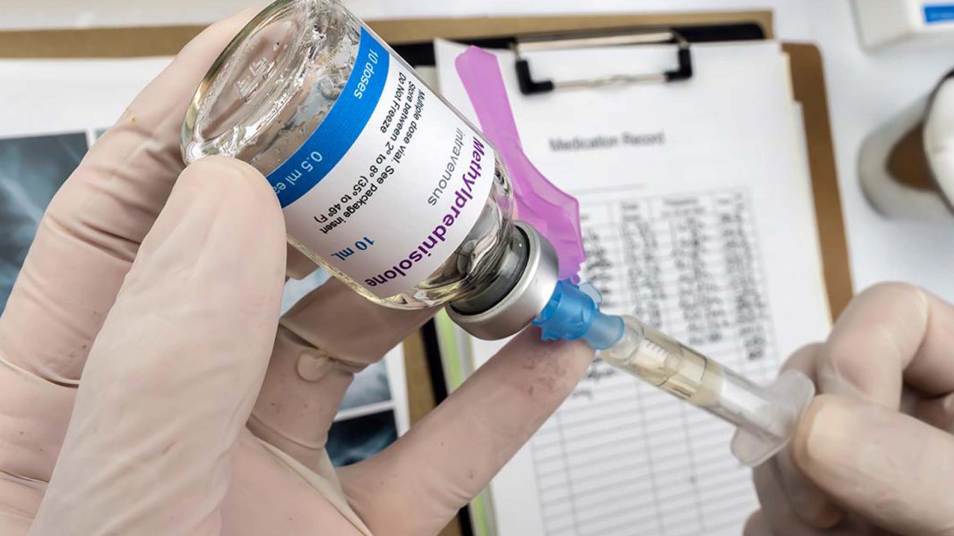
After the patient was placed on high-dose (500 mg per day) intravenous methylprednisolone for 3 days, she was switched to oral prednisone (60 mg per day) and her back pain resolved during the hospitalization. Methotrexate was added to her treatment regimen and she was successfully tapered off prednisone. A repeat abdomen/chest CT three months later showed that her aortitis had completely resolved; her ESR had also normalized. The patient continues to receive methotrexate, as well as IVIG for her SM, without any signs of relapse.
Beyond established etiologies of aortitis, doctors have reported large vessel vasculitis after infection with SARS-CoV-2, while others have linked systemic vascular dysfunction and endothelial cell injury in the setting of an aberrant cytokine response to the viral infection.. In fact, clinicians have described multiple vasculitic phenomena such as cutaneous vasculitis, Kawasaki-like disease spectrum, and pauci-immune thrombogenic vasculopathy in association with COVID-19. As probable explanations, they have proposed factors such as direct viral endotheliitis, complement-mediated vascular injury, and autoimmunity.
Because the patient developed aortitis very shortly after her SARS-CoV-2 infection, Dr. Homsi says he believes a hyperinflammatory state triggered by SARS-CoV-2 was likely behind her acute condition.
“Large vessel vasculitis, like aortitis after SARS-CoV-2 infection, carries a difficult diagnosis and treatment plan.”
Yamen Homsi, MD, MPH
“Large vessel vasculitis, like aortitis after SARS-CoV-2 infection, carries a difficult diagnosis and treatment plan,” Dr. Homsi says. “Fortunately, an immunosuppressant therapy including intravenous methylprednisolone has shown to be effective in treating the hyperinflammatory state in such patients.”