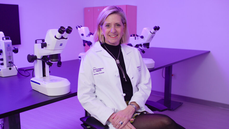
Disorders of the cornea are a leading cause of blindness worldwide. While surgical transplant of donor cornea is an effective treatment option, it remains out of reach for most patients. This large unmet need has helped to propel interest in developing cell-based regenerative therapies for blinding corneal diseases, says Shukti Chakravarti, PhD, a professor of ophthalmology and pathology at NYU Langone Health.
“A crucial step to developing such therapies is improving our understanding of corneal tissue development, as well as the interactions between corneal cell types,” Dr. Chakravarti explains.
Dr. Chakravarti and her research team are exploring the development of 3D organoids that could offer researchers a new investigative platform to better model the cellular complexity and functionality of the human cornea. They recently published preliminary results of their work in PNAS Nexus.
“Organoid technology has revolutionized the study of developmental stages and disease pathogenesis. We believe this new model provides a valuable 3D cell culture system for studying corneal disease mechanisms.”
Shukti Chakravarti, PhD
“Organoid technology has revolutionized the study of developmental stages and disease pathogenesis,” says Dr. Chakravarti. “We believe this new model provides a valuable 3D cell culture system for studying corneal disease mechanisms.”
Addressing Current Shortcomings
While some studies have investigated the single-cell transcriptomic composition of the human cornea, its organoids have not been examined extensively. Conventional 2D cell culture models often lack key information about tissue development and the consequences of interactions between cell types.
The researchers began their approach with single-cell RNA sequencing (scRNA-seq) of the central human cornea to determine its epithelial, stromal, and endothelial cellular landscape. They also performed scRNA-seq on human cornea organoids and compared the organoid single-cell transcriptome with that of the adult human cornea.
Modeling Corneal Diseases
In their report, Dr. Chakravarti and her team showcased the repertoire of cornea-like cells present in the organoid. As a whole, the organoid displayed features of a developing or a healing cornea.
“Our sequencing data show the presence of all three cell types in the organoids and, overall, their closer resemblance to developing rather than adult corneas,” says Dr. Chakravarti.
While the three major corneal cell types—epithelial, stromal keratocyte, and endothelial—are often investigated in single monolayer cultures, the 3D organoid system has already confirmed that interactions between all the three major layers are imperative for proper functioning of the cornea, she adds.
Future Applications
Based on these initial findings, the organoids offer a promising experimental system to model human diseases in tissue culture. While the 3D model appears to best conventional 2D cell culture models, further refinement and validation is still needed.
“Additional standardizations and sequencing of larger batches are needed for consistency. In the meantime, the cornea organoids may be used in functional studies of genes during early corneal development,” says Dr. Chakravarti.


