When a child arrives in an emergency department or urgent care with a suspected fracture, the location of that injury isn’t always immediately clear and the child is commonly evaluated with multiple X-rays. Patients and their families, however, have often told pediatric orthopedic surgeon Jody Litrenta, MD, about the stress of repeated imaging and long visits.
“I started to think that we could be more specific about the way we’re evaluating kids with injuries,” Dr. Litrenta says. In particular, she began wondering how to make the process more streamlined and effective.
In a study in the Journal of Pediatric Orthopaedics, she and colleagues at NYU Langone Health describe a simple radiographic protocol that successfully reduces the overall number of X-rays needed to thoroughly evaluate suspected fractures without missing any injuries.
“We don’t want to be the doctor that didn’t do enough. These results validate that a workup based on the protocol is appropriate.”
Jody Litrenta, MD
“We don’t want to be the doctor that didn’t do enough,” Dr. Litrenta says. “These results validate that a workup based on the protocol is appropriate.”
Playground Injuries
For their study, the researchers first conducted a retrospective chart review of 495 cases of suspected fracture in pediatric patients. “We wanted to see what kinds of injuries we were getting and what the current practice was,” she says.
Based on the data, the team created and distributed a simplified decision aid to help emergency room physicians and consulting residents think through when another X-ray is or isn’t necessary.
“There’s an idea that you need to X-ray the joint above and below the apparent injury. This is something that we do very often in a high-energy trauma when we’re concerned that in addition to a tibial shaft fracture, maybe there’s an injury in the knee or ankle,” Dr. Litrenta says. “But in kids with the lower-energy, playground injuries that we typically see here at NYU Langone, it’s generally safe to focus on one fracture area and not expand that.”
“In kids with the lower-energy, playground injuries that we typically see here at NYU Langone, it’s generally safe to focus on one fracture area and not expand that.”
The protocol’s three basic tenets include evaluating an injury with at most two contiguous areas of radiographs, limiting the screening for secondary fractures except for children with elbow injuries, and not repeating or obtaining additional imaging without a clear clinical concern.
X-Ray Reductions
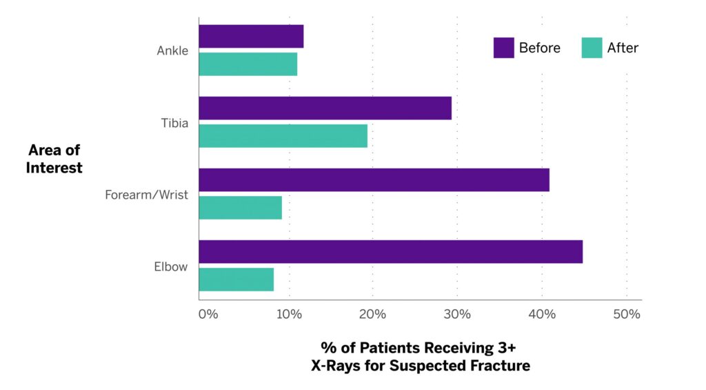
To assess the new protocol, the team evaluated its performance on an additional 333 pediatric injuries. Main outcome measures included the final diagnosis, total number of X-rays, number of anatomic areas imaged, visit length, and time for additional trips to radiology.
Crucially, the team found that the protocol still allowed for a comprehensive assessment that didn’t miss any injuries.
It also yielded a significant reduction in overall X-rays, especially for elbow injuries (Figure 1), and in X-rays for patients who had arrived with outside imaging. “Importantly, we saw that if those X-rays were negative, continuing on the hunt and sending the patient back for additional X-rays wasn’t helpful,” Dr. Litrenta says.

The researchers didn’t see a significant reduction in the length of emergency department visits, perhaps due to other limiting factors related to the administration of anesthesia and required observation times. Dr. Litrenta also cautions that car accident injuries and concerns about non-accidental trauma or abuse generally require more thorough X-ray evaluations.
For low-intensity injuries, though, the protocol proved its ability to reduce imaging with no loss in diagnostic rigor. “For the typical trauma that we see, it’s a good way to say that this is what you should start with and feel confident that you don’t have to escalate,” Dr. Litrenta says.


