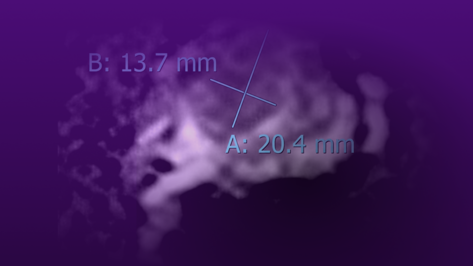


Inguinal lymph node dissection in penile cancer is often curative, but conventional open techniques are associated with a substantial complication rate. Minimally invasive, robotic approaches hold promise to greatly reduce the morbidity of this surgery. In addition, advancements in fluorescence lymphangiography have improved the intraoperative detection of pathologically involved nodes and lymphatic channels.
In this Case of the Month, we showcase the feasibility and efficacy of this robotic approach in an older male with an enlarged 1.6-cm left inguinal lymph node, which required a robotic-assisted bilateral inguinal lymph node dissection. We also highlight the role of fluorescence lymphangiography, which aided the removal of the sole site of metastasis and directed ligation of lymphatic channels to prevent lymphocele formation. The patient had no postoperative complications, and his wounds healed with excellent cosmesis.
Case Highlights:
- A man in his mid-70s underwent total penectomy and perineal urethrostomy creation for pT3, HPV-associated, G3 invasive basaloid squamous cell carcinoma of the penis.
- After a 1.6-cm right external iliac node tested negative for carcinoma and responded to antibiotics, a palpable 1.6-cm left inguinal lymph node developed.
- He underwent robotic bilateral inguinal lymph node dissection, with Firefly near-infrared lymphangiography guiding removal of 34 nodes.
- Pathology confirmed carcinoma in one left-sided lymph node with no extranodal extension.
- Early surveillance imaging demonstrated no evidence of residual or metastatic disease.
Patient Case
A patient in his mid-70’s presented to NYU Langone with a foul smelling, progressively worsening ulcer on his penis. Physical examination revealed a necrotic mass replacing the entire penile shaft, concerning for penile carcinoma. He underwent a total penectomy and perineal urethrostomy creation, with pathology confirming a pT3, HPV-associated, G3 invasive basaloid squamous cell carcinoma of the penis invading the corpora cavernosa, with negative surgical margins.
Initial surveillance CT imaging demonstrated an enlarged 1.6-cm right external iliac node, which was 1.2 cm prior to the penectomy. After his case was discussed at our multidisciplinary genitourinary tumor board, the consensus decision was to proceed with a percutaneous biopsy of the right external lymph node, which was negative for carcinoma.
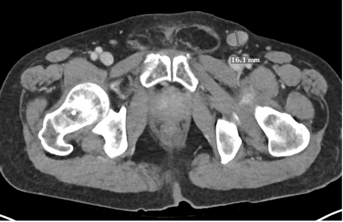
After a 4-week course of antibiotics, subsequent CT imaging demonstrated a decrease in the size of the right external iliac node to 1.2 cm, with a newly enlarged 1.6-cm left inguinal lymph node (Figure 1). This left inguinal lymph node was palpable and mobile on physical examination, with no other palpable inguinal lymph nodes.
Given the patient’s unilateral palpable lymph node and high-risk primary lesion, he was recommended to undergo a bilateral inguinal lymph node dissection.1 After thorough counseling, the patient agreed to proceed with robotic bilateral inguinal lymph node dissection.
Robotic Bilateral Lymph Node Dissection
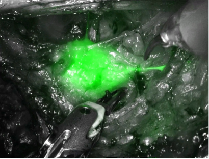
Patient Positioning and Access. The patient was placed in low lithotomy position. A half milliliter of 2 mg/kg indocyanine green was injected into the base of the prior penectomy site to facilitate lymph node identification during dissection (Figure 2).2 A line was drawn from the anterior superior iliac spine and pubic tubercle on one side. At 25 cm inferior to the midpoint of this line along the medial aspect of the thigh, a cut-down was performed to Scarpa’s fascia. Finger dissection was performed to create enough space for 8-mm robotic ports to be placed medial and lateral to this initial incision. A Hasson balloon port was placed through the initial incision and a 12-mm Airseal port was placed between the Hasson balloon port and the lateral robotic port.
Superficial Dissection. Using the medial border of the adductor longus muscle, the lateral border of the sartorius muscle, the superior border of the inguinal ligament, and the posterior border of fascia lata, the superficial lymph node packets were dissected, carefully sparing the saphenous vein. All visible lymphatic channels were ligated with a Vessel sealer or clips.
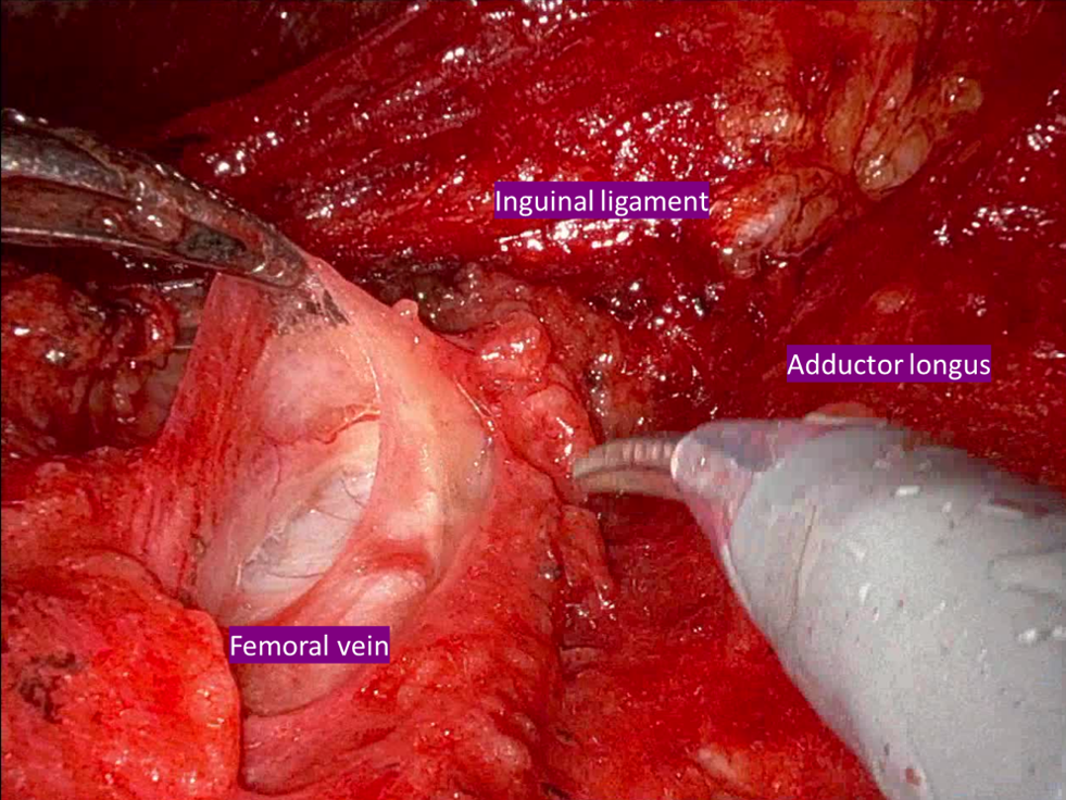
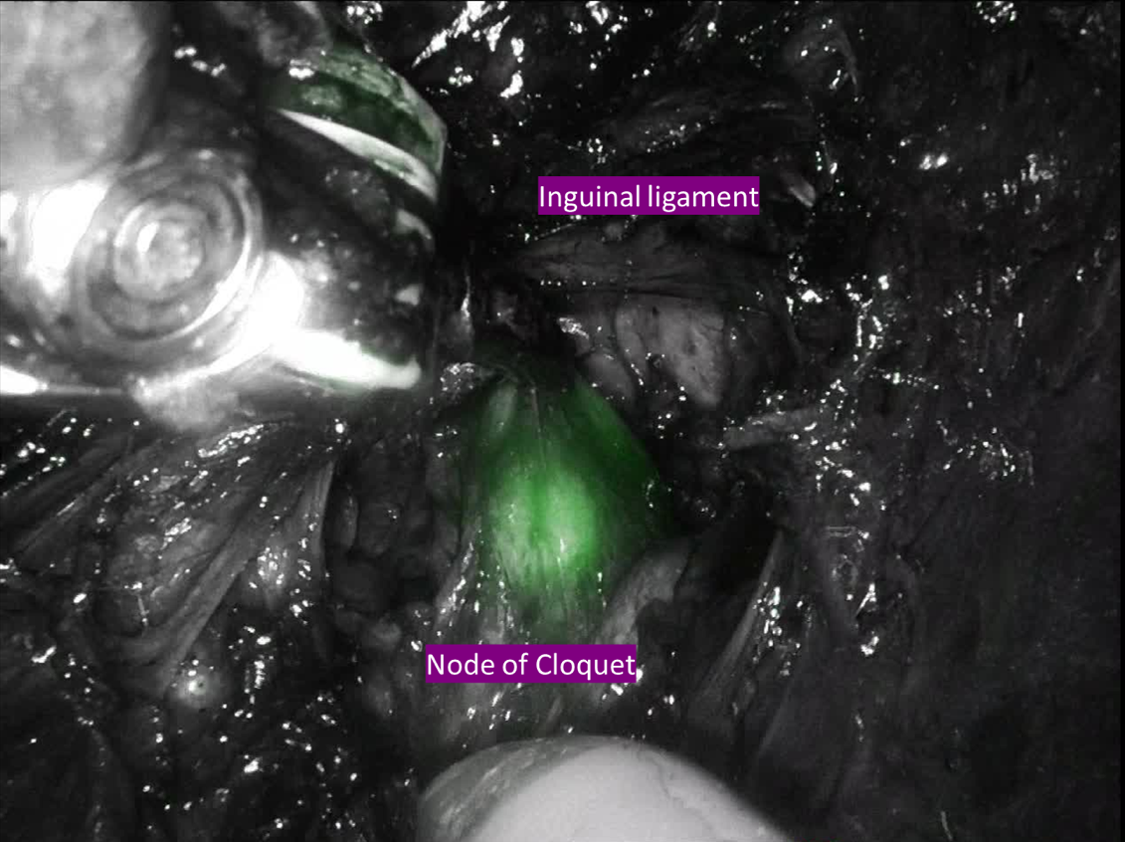
Deep Dissection. The fascia lata overlying the femoral vein was then incised (Figure 3). Staying medial to the femoral vein, the deep inguinal lymph node packet was dissected under the inguinal ligament up to the node of Cloquet (Figure 4).
Results. The total robotic console time was 167 minutes on the left side and 154 minutes on the right side. The patient was discharged home on postoperative day 1 with one Jackson-Pratt drain on each side. The drains were removed 6 weeks later, once the daily output of each drain was less than 30 milliliters.
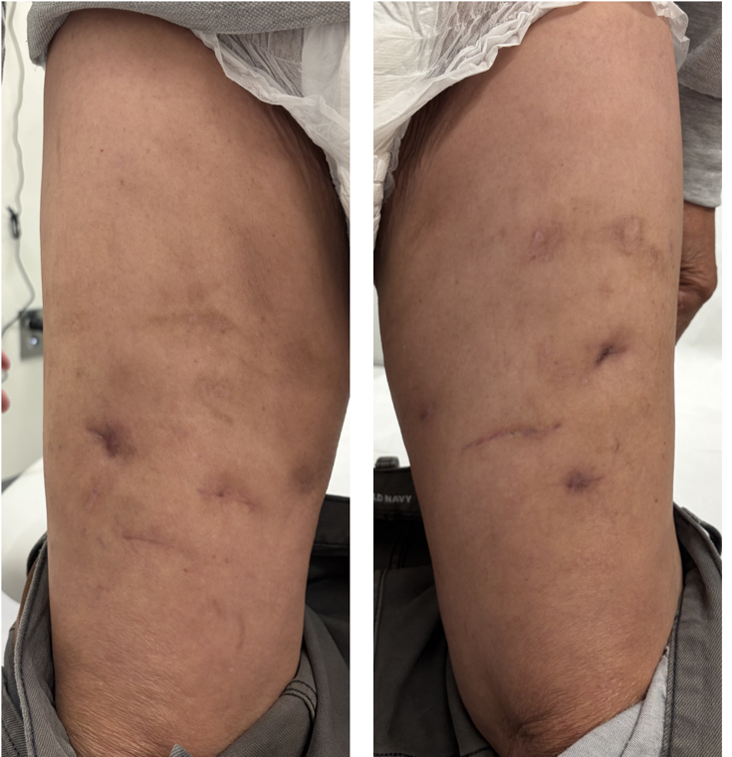
He had no postoperative complications and his surgical wounds healed well (Figure 5). Pathology demonstrated carcinoma in one of 19 left-sided lymph nodes (17 superficial and 2 deep)—with a tumor focus greater than 2 cm and no extranodal extension—and in none of 15 right-sided lymph nodes (12 superficial and 3 deep).
Surveillance. Given his pN1 disease, he was recommended to undergo monitoring as per NCCN guidelines, requiring physical examination, CT of the abdomen and pelvis, and a chest radiograph every 6 months for the first 2 years, then annually for years 3 and 4.1 Surveillance imaging 4 months after his inguinal lymphadenectomy demonstrated no new inguinal lymphadenopathy and no evidence of metastatic disease in the chest, abdomen, or pelvis.
Discussion
In the United States, about 2,260 new penile cancers will be diagnosed in 2026, accounting for less than 1 percent of all cancers.3 The surgical management of this rare disease often involves dissection of the inguinal lymph nodes, as they are the initial site of metastasis in almost all patients with invasive penile cancer. Therefore, inguinal lymph node dissection is critical for staging and may be curative in a significant portion of patients.
Traditionally, radical open inguinal lymph node dissection was the standard for managing inguinal nodes in penile cancer, but it was associated with a very high complication rate of 80 to 100 percent. The modified template, introduced by Catalona for patients with clinically negative nodes, significantly decreased the risk of complications and is still used today. More contemporary studies report a lower but still substantial complication rate of 42 to 57 percent. Common complications include skin edge necrosis, lymphedema, wound infection, and seroma formation.4 These can be long-term and debilitating for patients.
More recently, minimally invasive approaches, such as the robotic approach, have been introduced to further reduce the morbidity of this surgery. Early experience suggests that robotic surgery may shorten hospitalization, improve cosmesis, and potentially reduce lymphatic complications compared to open surgery.2,5 In addition, fluorescence lymphangiography, as first described at NYU Langone, improves the identification of pathologically involved nodes and lymphatic channels.2
This case demonstrates the advantages of the robotic approach in this setting. The pathologically involved lymph node and lymphatic vessels were easily identified on fluorescence lymphangiography, facilitating the removal of the sole site of metastasis and directed ligation of lymphatic channels to prevent lymphocele formation. A total of 34 lymph nodes were removed, an important quality metric as lymph node yield during inguinal dissection has been associated with overall survival in these patients.6 In addition, the patient was discharged home on postoperative day 1, he had no postoperative complications, and his wounds healed with excellent cosmesis. Early surveillance imaging demonstrated complete removal of his disease with no evidence of metastasis.
References
- National Comprehensive Cancer Network. NCCN Clinical Practice Guidelines in Oncology: Penile Cancer. Version 1.2026. Available at: NCCN.org. Accessed August 16, 2026.
- Bjurlin MA, et al. Urology. 2017;107:267. DOI.
- American Cancer Society. Key statistics for penile cancer. Accessed August 16, 2026. https://www.cancer.org/cancer/types/penile-cancer/about/key-statistics.html
- Spiess PE, et al. World J Urol. 2009;27(2):205-212. DOI.
- Nabavizadeh R, et al. Urol Oncol. 2023;41(1):1-14. DOI.
- Soodana-Prakash N, et al. Urol Oncol. 2018;36(10):471.e19-471.e27. DOI.