Referral Notes:
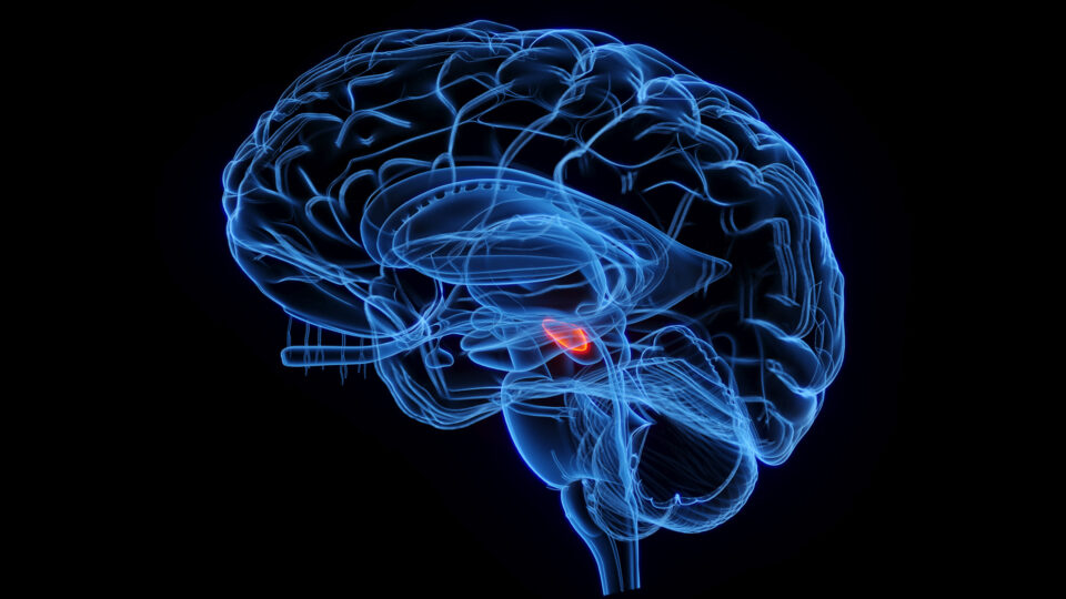
- The imaging agent used in fluorescence-guided glioma surgery, 5-ALA, has long been thought to cause the selective accumulation of the fluorophore PpIX in tumor cells, but technical limitations prevented testing this hypothesis.
- An NYU Langone-led study found no correlation between PpIX intensity and tumor cell concentration, but detected five distinct PpIX fluorescence patterns, distributed among extracellular and intracellular spaces.
- PpIX accumulated most avidly in cells identified as tumor-associated macrophages.
- These findings suggest PpIX fluorescence should not be relied on as the sole tumor marker in 5-ALA-guided glioma surgery, and that research on macrophages could reveal novel immunotherapeutic approaches for gliomas.
The only approved optical imaging agent for fluorescence-guided glioma surgery is 5-aminolevulinic acid (5-ALA), a pro-drug that metabolizes into the fluorophore protoporphyrin IX (PpIX). 5-ALA-guided surgery has been shown to enable more complete resections of tumors than conventional microsurgery. However, widely employed techniques for visualization of 5-ALA-induced PpIX fluorescence are limited by poor sensitivity, particularly at tumor margins.
In a study published recently in Nature Biomedical Engineering, a team led by NYU Langone Health researchers used a novel microscopy technique to improve the clinical detection of PpIX and gain a better understanding of its mode of action. Their work revealed several unexpected findings—including a tumor-independent mechanism, mediated by immune cells, for PpIX accumulation in human brain tumors.
“Our study shows how innovations in optics can advance our understanding of clinical fluorophores, and point toward new approaches to immunotherapy for gliomas.”
Daniel A. Orringer, MD
“Our study shows how innovations in optics can advance our understanding of clinical fluorophores, and point toward new approaches to immunotherapy for gliomas,” says neurosurgeon Daniel A. Orringer, MD, who led the study.
Pinning Down an Elusive Molecule
Approved by the European Medicines Agency in 2007, and by the U.S. Food and Drug Administration a decade later, 5-ALA has been used in a large number of glioma surgeries. The approach emerged from research showing that after patients ingested 5-ALA, their tumors fluoresced when exposed to light of certain wavelengths. Theoretically, that happened because PpIX was taken up preferentially by tumor cells. Yet, due to limitations in imaging technology, this hypothesis had never been tested empirically.
Dr. Orringer was inspired to do so, in part, by a phenomenon that he and others had observed: false positive fluorescence. “A lot of PpIX was ending up in tissue where there wasn’t tumor,” he recalls. “One of my interests is in developing technologies that enhance surgeons’ ability to visualize tissue on a microscopic level, and I wanted to see if we could get a better sense of which cells were actually concentrating PpIX.”
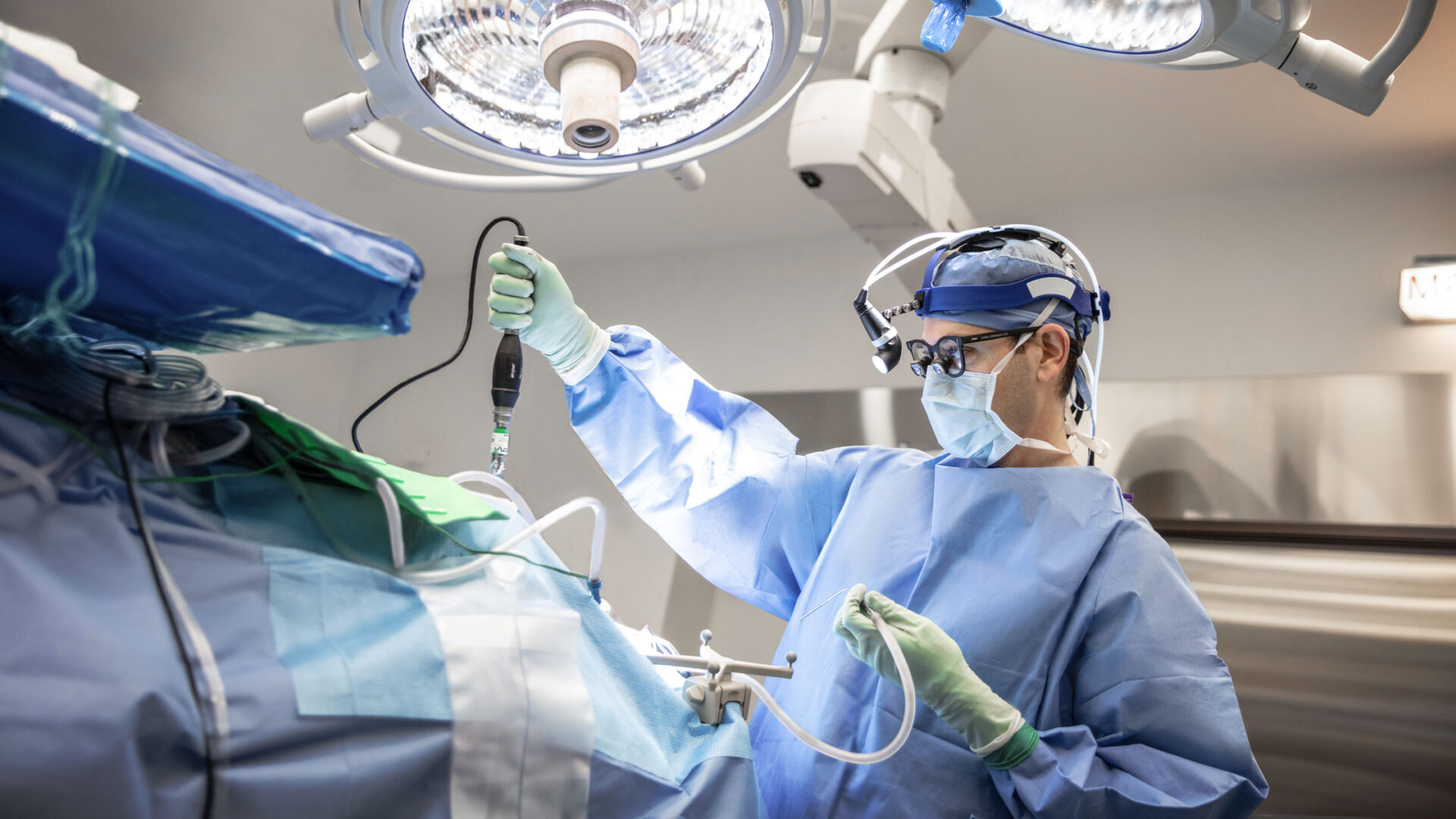
To that end, Dr. Orringer and colleagues took a system they had developed to distinguish tumor regions from normal tissue during surgery—intraoperative stimulated Raman histology (SRH) coupled with artificial intelligence-powered imaging analysis—and modified it. The revised system paired SRH with two-photon excitation fluorescence microscopy (TPEF) at the same spatial coordinates within each tissue specimen.
“We hoped that this bifunctional approach would allow us to localize PpIX in brain tissue with unprecedented precision,” he explains.
An Unsuspected Role for Macrophages
For the study, Dr. Orringer and his team examined fresh brain tumor specimens from 115 patients with high-grade gliomas. “The results were remarkable and surprising,” he says.
Contrary to the established hypothesis, the researchers found no correlation between PpIX intensity and concentration of tumor cells. Instead, semi-supervised clustering of TPEF images demonstrated five different patterns of PpIX fluorescence, distributed among extracellular as well as intracellular spaces.
“These scavenger immune cells were just gobbling up the dye.”
Further analysis, using spatial transcriptomics and metabolomics, revealed that PpIX accumulated most avidly in cells of myeloid origin, morphologically consistent with tumor-associated macrophages. “These scavenger immune cells were just gobbling up the dye,” says Dr. Orringer. “But we saw great variations in the abundance of macrophages both within the tumor and from patient to patient.”
Finding New Targets for Immunotherapy
These findings, in Dr. Orringer’s view, could have “profound implications” for the use of 5-ALA-guided glioma surgery. To begin with, they may help explain why previous research has shown a marginally higher risk of neurological deficits in patients who undergo such procedures, compared with those who receive conventional surgery: the technique can lead to the removal of healthy tissue whose glow falsely marks it as pathological.
That possibility makes relying on PpIX alone in glioma resection “a little dangerous,” he says. “My own surgical team takes a more conservative approach, combining fluorescence with histology to confirm our suspicion that a part of the brain contains tumor or is clear of tumor.”
The study could also point the way toward new approaches to glioma treatment. “We’re currently working to understand the biology of the macrophages that take up PpIX, and to determine whether they belong to an immunogenic or immunosuppressive sub-population,” Dr. Orringer says. “They may be of interest in developing therapies that target the immune system to fight these terrible malignancies.”